


28, Feb 2024
Minister Amber-Jade Sanderson’s Hyderabad Mission Strengthens Healthcare Collaboration between Western Australia and India
 Hyderabad, February 28, 2024: Minister for Health and Mental Health, Amber-Jade Sanderson embarked on a mission to India to strengthen WA-India links across the healthcare, medical, and life sciences sectors. Leading a health skilling and business delegation, the Minister has concluded her highly productive mission to Hyderabad, spanning from February 25 to February 27, 2024. The delegation actively participated in numerous engagements, featuring representatives from industries including health and life sciences, medical technology, digital health, and medical device companies, biotech, and pharmaceutical companies.
Attracting healthcare workers is a key priority for the Western Australian government, with forecasts predicting WA will need an additional 5,000 FTE doctors and nurses by 2033. During the mission, Minister Sanderson successfully promoted employment opportunities in Western Australia’s healthcare sector, with a specific focus on professions including nurses, doctors, and aged care workers. She held crucial meetings with esteemed dignitaries, including Hon. Damodar Raja Rajanarasimha, Minister of Health, Medical & Family Welfare, Science and Technology, to foster discussions on collaboration and investment in healthcare.
The Health Skilling Symposium, held in collaboration with Rainbow Hospital and Telangana Overseas Manpower Company (TOMCOM), facilitated meaningful discussions and initiatives aimed at enhancing skills and capacity-building efforts in the healthcare domain. Minister Sanderson’s participation at BioAsia 2024 provided a platform to showcase Western Australia’s capabilities in health and life sciences, fostering connections and exploring potential collaborations.
During the mission, Minister Sanderson held important meetings with distinguished figures including Hon. Sri Damodar Raja Narasimha, Minister for Health, Medical & Family Welfare, Science and Technology, Telangana and bilateral meetings with Hon. Anumula Revanth Reddy, Chief Minister, Telangana, and Hon. Duddilla Sridhar Babu, Minister for Information Technology, Electronics & Communications; Industries & Commerce and Legislative Affairs, Government of Telangana, strengthened ties and identified areas for cooperation in healthcare.
Minister Sanderson highlighted Western Australia’s vibrant healthcare ecosystem, showcasing advancements in healthcare, research, and development, and fostering interest in potential investments. She reiterated the government’s commitment to supporting skilled migrant workers through initiatives like the Skilled Migrant Job Connect Program. The Western Australian Premier’s University Scholarship was showcased to support students pursuing healthcare-related courses, further bolstering the skilled workforce in the region.
Minister Sanderson was also delighted to announce the signing of a collaboration agreement between ECU and Charkos Global, aimed at facilitating the education and migration of nurses. The partnership will facilitate supporting Indian nurses through transition courses at ECU including the award-winning International Adaptation Program (winner of AAUT INTERNATIONAL Teaching Excellence Award in 2022). ECU will also partner with Charkos Global to accredit specialist post-graduate courses for nurses in India working in pediatrics, mental health, and oncology nursing. We expect the first Indian nurses to come to ECU in August this year.
A significant highlight of the mission was Minister Sanderson’s keynote address at the 21st Bio Asia 2024 Summit, a premier life-sciences and healthcare forum in Asia where innovation, data, and artificial intelligence converge to redefine possibilities. This two-day conference catalyzed furthering the ties between Western Australia and India. At this event, Western Australia partnered with Bio Asia as a “Global Partner” which allowed us to showcase Western Australia’s complementarity with the state of Telangana in the sectors of Healthcare and Life Sciences.
Reflecting on the engagements in Hyderabad, Minister Sanderson remarked, “Through our mission to Hyderabad, we’ve forged invaluable connections and laid the groundwork for transformative collaborations between Western Australia and India. Our shared commitment to advancing medical innovation and fostering skilled workforce exchange lays a solid foundation for addressing healthcare challenges and driving economic prosperity. I’m immensely proud of the outcomes achieved and look forward to the continued growth and prosperity that these partnerships will bring.”
- 0
- By Rabindra

22, Feb 2024
Physiotherapy Heals Wounds
By Dr S.Senthilkumar,
Department of hand and General Surgical Physiotherapy,
Saveetha College of physiotherapy,
Thandalam
Pressure sores and wound ulcers are debilitating complication of diabetic patients as well as other patients who met with road traffic accidents. Delay in healing of wounds concerns every patient and affects their normal activities of daily life. Various drug regimens were used to promote wound healing. Iodine, saline and anti bacterial ointments facilitate wound healing. Physiotherapy also plays an important role in promoting wound healing. Low intensity laser therapy, Ultrasound therapy and ionotophoresis indeed help in wound healing.
Laser therapy
Low intensity laser therapy is recently used for the treatment of wounds. Laser accelerates the process of healing of wounds. Laser facilitates wound healing by chemotactic activity, vascular changes and chemical mediators. Laser therapy enhances tissue proliferation and thus facilitates wound healing caused due to burns, pressure sores, diabetic ulcers and pressure sores. Chronic and acute ulcers treated effectively by laser therapy using direct contact or grid method.
Ultrasound therapy
Ultrasound therapy enhances wound healing in all stages of repair. It increases permeability of lysozomes membrane to clear debris from wound area. It also induces calcium ions into the wound causing mobility of fibroblasts to form scars while myofibroblasts contracts wound. Ultrasound therapy also increases the tensile force enhancing strength and elasticity of the fibres. Ultrasound effectively heals pressure sores
Ionotophoresis:
Wound healing also promoted by using the principles of ionotophoresis. Zinc oxide is used in ulcers to enhance the healing of wound.
22, Feb 2024
Exploring the Role of Pelvic Wands in Pelvic Floor Rehabilitation
By Dr. Suriya. N
MPT ( obg), Tutor, Saveetha College of Physiotherapy,
saveetha institute of medical and technical sciences, Tamil Nadu, India.
Introduction:
Pelvic floor dysfunction is a prevalent condition affecting individuals of all ages and genders worldwide. The pelvic floor muscles play a crucial role in supporting the pelvic organs, maintaining continence, and facilitating sexual function. Dysfunction in these muscles can lead to various issues such as urinary incontinence, pelvic pain, and sexual dysfunction. Pelvic floor rehabilitation aims to restore strength, flexibility, and coordination to these muscles. One tool that has gained recognition in this field is the pelvic wand. In this article, we delve into the significance of pelvic wands in pelvic floor rehabilitation and how they are used in therapy.
Understanding Pelvic Wands:
A pelvic wand, also known as a pelvic floor wand or trigger point wand, is a specialized tool designed to assist in pelvic floor muscle rehabilitation. It typically consists of a slender, wand-like device made from medical-grade materials such as silicone or plastic. At each end of the wand, there may be various features such as rounded knobs, ridges, or curves, which serve different purposes during therapy.
Role of Pelvic Wands in Pelvic Floor Rehabilitation:
Trigger Point Release:
Pelvic wands are often used to perform trigger point release therapy. Trigger points are hypersensitive areas within the muscles that can cause pain and dysfunction. By applying gentle pressure to these trigger points using the wand, therapists can help alleviate muscle tension and discomfort, promoting relaxation and improved function of the pelvic floor muscles.
Myofascial Release:
Myofascial release is a technique used to release tension and tightness in the fascia, the connective tissue surrounding muscles. Pelvic wands can be effective tools for performing myofascial release in the pelvic region. Therapists use the wand to apply controlled pressure to specific areas of the pelvic floor, helping to stretch and release tight fascia, thereby improving mobility and reducing pain.
Pelvic Floor Muscle Strengthening:
In addition to releasing tension, pelvic wands can also be utilized to facilitate pelvic floor muscle strengthening exercises. By incorporating the wand into exercises such as pelvic floor contractions (Kegels), therapists can provide tactile feedback and enhance muscle awareness, helping individuals better engage and strengthen their pelvic floor muscles.
Biofeedback:
Some pelvic wands are equipped with sensors or markings that provide biofeedback to the user. This real-time feedback allows individuals to monitor their pelvic floor muscle activity and ensure they are performing exercises correctly. By using a biofeedback enabled pelvic wand, individuals can optimize their pelvic floor rehabilitation efforts and track their progress over time.
Self-Treatment:
One of the key advantages of pelvic wands is that they enable individuals to perform self-treatment in the comfort of their own homes. With proper instruction from a pelvic floor therapist, individuals can learn how to use the wand safely and effectively to manage their symptoms and improve pelvic floor function between therapy sessions.
Conclusion:
Pelvic wands are valuable tools in pelvic floor rehabilitation, offering a range of benefits including trigger point release, myofascial release, muscle strengthening, biofeedback, and self-treatment. When used under the guidance of a qualified pelvic floor therapist, pelvic wands can help individuals alleviate symptoms, improve pelvic floor function, and enhance overall quality of life. As awareness of pelvic floor dysfunction continues to grow, pelvic wands are likely to remain an essential component of comprehensive pelvic floor rehabilitation programs.
22, Feb 2024
Diabetes Surgery Successfully Performed on A 41-year-old Woman with Uncontrolled Diabetes
Mumbai: A team headed by Dr Ramen Goel, Consultant Diabetes and metabolic surgeon, Wockhardt Hospitals, Mira Road achieved a breakthrough by successfully conducting a diabetes surgery on a 41-year-old woman who has been battling uncontrolled diabetes for over a decade. The patient also suffered from other health issues like high blood pressure, high cholesterol, and thyroid problems, which greatly affected her overall well-being. This surgical intervention has been gaining traction for the last 1 year and significantly reduces the chances of developing complications associated with diabetes, such as diabetic retinopathy, brain stroke, kidney disease, and cardiac and nerve problems, all of which contribute to higher mortality rates in our country. Remarkably, this surgery improves blood sugar levels, and aids quick recovery and the patient has now completely stopped taking diabetes medication and has seamlessly resumed her daily activities.
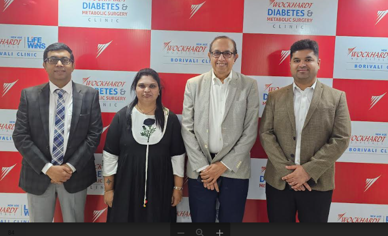
Patient Mrs Kavita Mahesh, a 41-year-old resident of Mira Road, weighing 84.5 kgs had uncontrolled diabetes for 10 years besides high blood pressure, high cholesterol, and thyroid. Additionally, her father has kidney problems. Despite taking the medication regularly, her blood sugar levels remain uncontrolled. In November, the patient approached Wockhardt Hospitals, Mira Road, and sought assistance due to her inability to control her blood sugar levels and the presence of high cholesterol.
Dr. Ramen Goel, Consultant Diabetes and Metabolic Surgeon, Wockhardt Hospitals Mira Road said, “The prevalence of diabetes mellitus is rapidly growing, affecting a staggering number of people in the country. Despite extensive awareness campaigns and lifestyle modifications, over 77% of people with diabetes, have failed to achieve proper control over their condition (indicated by HbA1c levels above 7). As a result, patients with uncontrolled diabetes face an increased risk of developing macrovascular complications such as cardiovascular diseases (including heart attacks) and brain strokes, as well as microvascular complications like peripheral neuropathy and nephropathy. The impact of this disease extends beyond a reduced lifespan, with an estimated millions of lives in India being lost due to direct causes related to diabetes. Hence, diabetes surgery can be a boon for patients with high HBA1c levels that help to regularise average blood sugar levels within 2-3 months.”
“Diabetes surgery is a laparoscopic gastrointestinal procedure that reroutes the stomach and the intestines. In India, gastric bypass and sleeve gastrectomy are commonly performed metabolic surgeries. While these surgeries were originally intended for weight loss, they can now be offered to patients with a BMI of 27.5 who have uncontrolled diabetes. The surgeries work by changing gut hormones, and gut bacteria, and limiting food intake, resulting in improved insulin sensitivity, increased insulin secretion, and better glycemic control. Its purpose was to redirect food to the latter part of the intestine, which secretes a hormone called GLP-1. This hormone reduces insulin resistance and stimulates increased insulin production from the pancreas. The surgery was done on February 6th, and the patient was discharged on February, 9. The surgical procedure typically takes around one hour, and patients are allowed to drink water and walk within 6-8 hours after surgery. She is completely off diabetes medicines 1 week after discharge and her HAB1C levels have already dropped by 10% The recovery process after metabolic surgery is relatively quick and the blood sugar levels start improving within the first two days after surgery. In most cases, injectable insulin can be discontinued within 2-7 days post-surgery,” said Dr Goel.
Individuals who undergo diabetes surgery experience glycemic resolution, and their blood sugar levels return to normal, and most no longer require medication. “Most patients continue to be free from diabetes medication even a decade after the surgery. This sustained maintenance of normal blood sugar levels has a remarkable impact on reducing complications associated with diabetes. After diabetes surgery, there is a reduction in diabetic retinopathy (eye problems), diabetic kidney disease, and diabetic nerve problems. There is also a decrease in diabetes-related heart attacks and deaths. The achievement of glycemic control and the avoidance of complications are crucial factors in managing diabetes effectively,” underscored Dr Ramen Goel.
Wockhardt Hospitals, Mira Road has taken a pioneering step by introducing metabolic surgery in North Mumbai. This innovative procedure goes beyond traditional weight loss surgeries, as it is specifically designed to treat diseases such as diabetes and hypertension and transform lives for the better. This commitment to improving patients’ overall health reflects the Hospital’s dedication to providing holistic healthcare solutions that go beyond mere symptomatic treatment; highlighted Dr. Pankaj Dhamija, Centre Head, Wockhardt Hospitals, Mira Road.
“Dr Goel transformed my life by performing the surgery which not only improved my blood sugar levels but also hopefully saved me from dangerous complications. Before the surgery, I was constantly battling high blood sugar levels despite multiple medications and strict dietary control. It felt like an endless cycle of frustration and despair. Dr. Goel’s expertise in diabetes surgery offered a glimmer of hope for a better future. It not only drastically reduced my blood sugar levels but also provided me with newfound freedom and peace of mind. My quality of life has significantly improved and I now enjoy activities that were once lost to chronic fatigue due to uncontrolled diabetes. He explained how reducing excess weight through metabolic surgery can lead to improvements in blood pressure, cholesterol levels, and overall cardiovascular health by reducing the risk of complications. I am forever indebted to Dr Goel and his team,” concluded the patient Mrs Kavita Mahesh
22, Feb 2024
New genetic therapy could be a gamechanger for Motor Neurone Disease and frontotemporal dementia
New Delhi, 22 February 2024: Macquarie University neuroscientists have developed a single-dose genetic medicine that has been proven to halt the progression of both motor neurone disease (MND) and frontotemporal dementia (FTD) in mice – and may even offer the potential to reverse some of the effects of the fatal diseases.
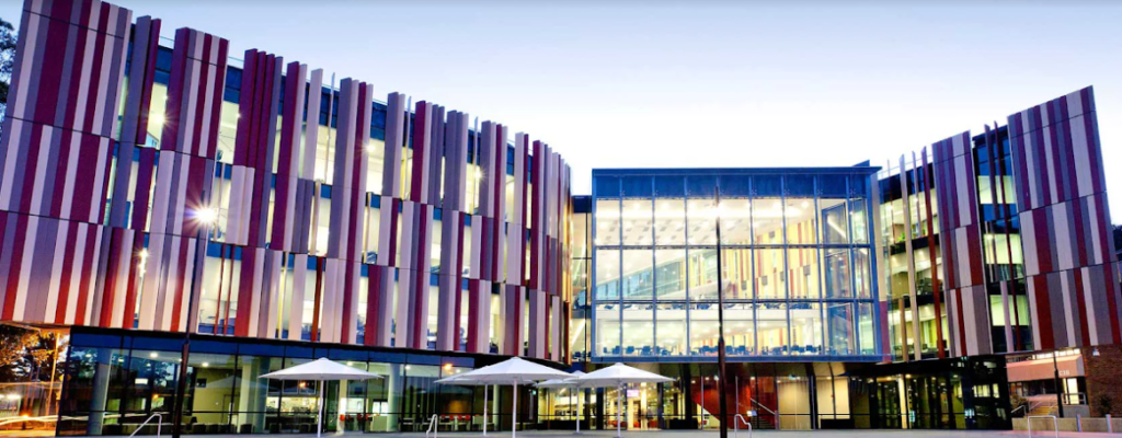
It may also hold opportunities for treating more common forms of dementia, such as Alzheimer’s disease. The new treatment, dubbed CTx1000, targets pathological build-ups of the protein TDP-43 in cells in the brain and spinal cord.
Cells like neurons produce TDP‑43 naturally, and it is important for their healthy function. Under certain conditions, it accumulates in the wrong part of the cells, clogging them and preventing them from working properly. For the past 15 years, a Macquarie University research team led by Professor Lars Ittner has been looking for the causes of this pathological build-up of TDP-43, along with ways to clear the blockages and prevent them from forming in the first place.
The team’s latest findings are published in the prestigious international neuroscience journal, Neuron.
Professor Ittner says their research has furthered their understanding of MND and FTD and their causes.
“We discovered for the first time that where there is pathological TDP-43, there is also an increase in a second protein, 14‑3‑3,” elaborated Professor Ittner.
“The two proteins interact, resulting in these build-ups in the cells. From this, we were able to isolate a short peptide that controls this interaction, and that’s what we used to create CTx1000. When we administered it in the lab, it dissolved the build-ups, tagging TDP-43 proteins for recycling by the body, and prevented new ones from forming. Importantly, CTx1000 targets only pathological TDP-43, allowing the healthy version of the protein to be produced and go about its work unhindered.”
This makes CTx1000 incredibly safe, and Professor Ittner says they have seen no adverse effects in their studies. “We have great hopes that when this progresses to human trials, it will not only stop people from dying from both MND and FTD, but even allow patients to regain some of the lost function through rehabilitation.”
Professor Yazi Ke, lead author of the new paper, says the milestone is particularly exciting for her because the original discovery was made in her early postdoctoral days. “This spans more than a decade of work, and goes from discovery all the way through to a potential treatment,” she says.
“In lab conditions, we saw CTx1000 stop MND and FTD from progressing even at very advanced stages, and resolving the behavioural symptoms associated with FTD. We have great hopes that when this progresses to human trials, it will not only stop people from dying from both MND and FTD, but even allow patients to regain some of the lost function through rehabilitation.”
Research Fellow Dr Annika van Hummel says they covered a variety of mutations in the TDP genes during lab testing.
“We wanted to prove beyond a doubt that this would work in different situations, and it resulted in clear improvements in both symptoms and brain pathology,” she says.
“The fact that it was effective both before and after the onset of symptoms makes it particularly exciting. While we are initially concentrating on MND and FTD, but about 50 per cent of cases of Alzheimer’s also show TDP pathology, so it’s possible that in the future this treatment could be translatable to other neurodegenerative conditions.”
A desperate need for treatment options
Also known as amyotrophic lateral sclerosis (ALS), MND causes the progressive loss of the neurons that allow the brain and spine to communicate with the muscles. In its early stages, patients experience muscle weakness, but as the disease progresses, they gradually lose the ability to walk, speak, swallow and breathe unaided. Most people with MND die within two to five years of diagnosis.
FTD is one of the rarer forms of dementia, but it is the second-most common form in people younger than 65. Actor Bruce Willis was diagnosed with FTD in 2023. It does not always have obvious physical symptoms, but it results in cognitive decline coupled with behavioural symptoms including anxiety, loss of inhibition, personality change and impaired judgment. Patients may live for more than 10 years after diagnosis, but it is ultimately fatal.
While there is a genetic therapy showing promise for one form of familial MND, there are few treatments available for the sporadic MND that makes up 90 per cent of all cases. Of those, the most effective can only extend a patient’s life by up to five months. All require frequent doses, and some come with side effects that are difficult to cope with.
There is currently no treatment for FTD.
Macquarie University Professor of Neurology, Professor Dominic Rowe, is one of Australia’s leading MND clinicians and researchers.
“I am currently treating more than 200 people with MND, but only five of them with a specific type of familial MND are currently able to access genomic therapy,” Professor Rowe says.
“Treatment like that doesn’t exist for anyone else. For most patients, all we can do is provide good quality, multidisciplinary care. This new research is incredibly promising in slowing the progression of MND and FTD for the vast majority of our patients. I’m extremely hopeful that it will soon be available to our patients at the Macquarie University Hospital MND Clinic.”
The team at Macquarie University hopes to be able to begin in-human trials of CTx1000 within two years, and they have already received a pre-clinical grant of $1.2 million from FightMND to assist with the process.
The therapy is one of the key discoveries being championed by Celosia Therapeutics, a Macquarie University spin-out company formed in 2022 to help bring groundbreaking work of the University’s neuroscientists from the lab to patients.
Testing new therapies and bringing them to waiting patients is incredibly expensive – in this case, the cost is an estimated $22 million to get to human trials.
“There is such a desperate need for new drugs for neurodegenerative diseases like MND, and this is an exciting new therapeutic with enormous potential,” Dr Hamdorf says.
“MND is difficult to diagnose because there is no conclusive test. It’s a process of elimination that can take up to 12 or 18 months, by which time, the disease has progressed significantly. We have the potential for CTx1000 to be a one-shot treatment that can help people even at late stages of the disease, then it would quite literally be a light at the end of the tunnel that patients and their families just don’t have now.”
This research was funded by grants from the National Health and Medical Research Council and MND Australia.
Professor Lars Ittner is the Director of the Dementia Research Centre and Professor in the Department of Biomedical Sciences of the Faculty of Medicine, Health and Human Sciences at Macquarie University.
Professor Yazi Ke is the Deputy Director (Operations) of the Dementia Research Centre and Professor in the Department of Biomedical Sciences of the Faculty of Medicine and Health Sciences at Macquarie University.
Dr Annika van Hummel is a Research Fellow at the Macquarie University Dementia Research Centre.
Professor Dominic Rowe is the Foundation Professor of Neurology in the Faculty of Medicine and Health Sciences at Macquarie University, Chairman of Macquarie Neurology, and Consultant Neurologist at Macquarie University Hospital.
22, Feb 2024
Thyroid Eye Disease: more to it than meets the Eye
Authored by Dr. Saptagirish, HoD Orbit and Oculoplastics, Dean,
Sankara Eye Hospital, Bangalore
Thyroid Eye Disease (TED) is mostly caused by the immune system targeting the muscles and tissues that surround the eyes, which causes irritation and an array of ocular symptoms. In a study conducted over 18 months in 2020, it was found that 34.7% patients with thyroid dysfunction having TED and 4:1 ratio of women to men with TED. With the advancement of AI and technology, it is much easier to diagnose TED and provide much more advanced treatment that will help in the speedy recovery of the patient. People need to be conscious of these indications and get help as soon as possible to prevent them from getting worse. Failure to do so may lead to complications, including potential vision loss.
Key Clinical Features of Thyroid Eye Disease:
Protrusion of Eyes (Proptosis): One prominent indication entails the protrusion or bulging outwards of one or both eyes due to swelling and inflammation affecting the eye muscles along with adjacent tissues.
Double vision: TED can induce restriction of one or more muscles that help rotate the eyes in different directions, thereby leading to double vision medically termed diplopia.
Ocular Discomfort and Pain: Individuals afflicted with TED often experience discomfort accompanied by dull aching pain within their affected eyes coupled with sensations akin to grittiness, irritation and foreign body sensation.
Swelling and Inflammation: Swelling alongside inflammation in tissues surrounding the eyeballs leads to redness, pain and discomfort
Dry eyes: Reduced production of tears, inability to close eye completely and increased visualization of white part of the eye leads to dryness
Cosmetic disfiguration: bulging of one or both eyes, staring look, inability to close eyes completely and eyelid lag can result in unsightly appearance aesthetically displeasing to some individuals.
Systemic manifestations: Weight loss, palpitations, increased heart rate, heat intolerance and generalised apprehension and anxiety should alert one on presence of hyperactive thyroid hormones. Ocular manifestations may precede or follow systemic manifestations in a majority of individuals.
Dietary Considerations
When managing TED closely paying heed towards eating habits emerges as beneficial thus certain considerations worth pondering include:
Foods that should be avoided include processed foods, as they often contain high levels of sodium which can contribute to water retention and worsen eye swelling. Excessive intake of sugary foods should be restricted as it may lead to inflammation in the body. It is always recommended to follow a low-sugar diet to help manage symptoms.
Selenium supplements: Dietary sources like onions, garlic, meat, seafood, shellfish, eggs and cereals or selenium supplements prescribed by a medical practitioner have beneficial effects in controlling symptoms and progression in mild TED due to its antioxidant properties.
Addictions: Smoking and alcohol are substances that have the potential to exacerbate eye discomfort and disrupt sleep patterns associated with Thyroid Eye Disease (TED). Therefore, it is advisable to gradually reduce and stop consumption of these substances altogether.
To take precautions against TED, regular medical check-ups are essential. By scheduling routine appointments with healthcare providers, individuals can monitor their thyroid function and assess the progression of TED more effectively.
Lubricants: The use of lubricating eye drops can also alleviate dryness and increase overall comfort experienced by some individuals.
Adequate sleep plays a significant role in promoting overall well-being while alleviating uncomfortable symptoms such as eye discomfort. It’s important to prioritize quality rest by ensuring sufficient hours of uninterrupted sleep each night.
Furthermore, stress management techniques prove beneficial for managing autoimmune conditions related to TED. Adopting practices like meditation or yoga helps individuals cope better with stressors that could potentially worsen their condition over time.
Understanding Thyroid Eye Disease requires awareness of its symptoms alongside implementing supportive dietary choices and taking the necessary precautions mentioned above. This includes seeking personalized advice from healthcare professionals regarding tailored treatment plans. Finding a balance between medical guidance, and lifestyle modifications allows people affected by this condition, to improve their overall well-being effectively, and successfully manage its impact on ocular health.
• Indian Journal of Ophthalmology: Clinical profile of thyroid eye disease and factors predictive of disease severity
• National Library of Medicine: Selenium supplementation in thyroid associated ophthalmopathy, an update
22, Feb 2024
Uptick In Respiratory, and Cardiac Problems Due To Poor Air Quality In Delhi
Delhi:- As a result of poor air quality, many residents in the capital are experiencing respiratory and cardiac issues. Instances of cardiac arrest, stroke, and gastric problems have risen significantly. It is crucial to refrain from going outdoors during periods of low air quality or to wear a mask as a precaution.
“With levels of particulate matter (PM) reaching hazardous levels, the impact on respiratory and cardiac health is becoming increasingly evident. There is a spike in cases of asthma, bronchitis, and other respiratory illnesses, while individuals with pre-existing heart conditions are experiencing exacerbated symptoms. The high levels of pollutants in the air can lead to inflammation in the lungs and heart, putting individuals at a higher risk for cardiovascular diseases. There has been a notable rise in the number of patients seeking medical care for a variety of health concerns, including respiratory issues like asthma, chronic coughs, colds, viral infections, and abdominal distress. This increase has also been observed in cases related to cardiac conditions. People come with signs and symptoms such as coughing, wheezing, and shortness of breath, which are often exacerbated by pollutants. Children and the elderly population are particularly vulnerable to these effects due to weakened immune systems or respiratory function. Air pollution has been linked to cardiovascular problems such as heart attacks and strokes. last 2 month 200 cases spread across all age group, but mainly old age and adults with comorbidities like diabetes and heart disease.” Said Dr Dixit Thakur, Pulmonologist, Apollo Spectra Delhi, Chirag Enclave.
Dr Thakur Added, “ The high levels of pollutants in the air have been linked to respiratory issues such as asthma, chronic obstructive pulmonary disease (COPD), and bronchitis, as well as an increase in cardiovascular diseases. The presence of smog can cause direct irritation to the lungs and throat, especially impacting individuals with pre-existing respiratory issues. Common issues include upper respiratory problems such as nasal congestion, coughing, wheezing, difficulty breathing, and a sore throat. Individuals may also experience chest discomfort and fever. However, the majority of these symptoms are mild and typically resolve on their own. Urgent action must be taken to address this pressing issue and improve air quality in Delhi for the well-being of its residents.”
Investing in high-quality air purifiers equipped with HEPA filters can significantly improve indoor air quality and help combat the effects of pollution in Delhi. Limit outdoor activities during peak pollution hours, typically early morning and late evening when the pollutant levels are at their highest. Opt for indoor exercises or activities during these times to reduce exposure to toxic air pollutants and protect your respiratory health in the long run. Avoiding venturing out of the house or wearing a mask can help to keep the lungs and heart healthy,” Concluded Dr Thakur.
21, Feb 2024
Nestlé Health Science launches Resource Activ, the ‘New-Edge’ nutritional solution for active millennials
In line with its purpose of unlocking the power of food to enhance quality of life for everyone, today and for generations to come, Nestlé India has launched Resource Activ, a multi-benefit, high-protein supplement designed to cater to the wellness needs of active millennials. With its unique ‘New Edge Formula’, Resource Activ has high quality proteins for muscles, enriched calcium and vitamin D for bone health and hyaluronate for skin health. It also contains fiber and immune nutrients. Resource Activ seeks to address the nutritional need gaps of the millennials through supplementation.
Despite the rapidly growing protein supplement market, there is a need for offerings tailored for individuals. Studies suggest that after the age of 30, there is involuntary loss of muscle mass. It also leads to reduced bone density, tissue breakdown and signs of aging like loss of skin hydration and elasticity. Resource Activ is customized to fulfill the nutritional needs of this age group, aiding muscle health, energy, bone health and skin health. Consumers can enjoy Resource Activ with water or milk in a delicious vanilla biscuit flavour.
Talking about the launch, Mr. Suresh Narayanan, Chairman and Managing Director, Nestlé India said, “Nestlé India has been on a continuous journey to add value to the lives of its consumers through powerful brands and products. We are happy to launch Resource Activ, for active adults, including millennials. Resource Activ is an output of extensive research and is backed by the assurance of Nestlé’s quality and safety. We are confident that the brand would be able to supplement the nutritional needs of the active millennials and help them lead an active life.”
Resource Activ will be available on ecommerce and across 11 cities at national pharmacies and local chemists in Delhi NCR, Kolkata, Mumbai, Pune, Hyderabad, Bangalore, Chennai, Coimbatore, Kochi, Kozhikode, and Thiruvananthapuram.
Nestlé Health Science is a global leader in the space of nutritional science. It focuses on advancing the therapeutic role of nutrition to change the course of health management for consumers, patients, doctors, nurses and its partners in healthcare. The key products include RESOURCE High Protein, PEPTAMEN, OPTIFAST, RESOURCE Diabetic, RESOURCE Fiber Choice, THICKEN UP CLEAR, RESOURCE Renal and RESOURCE Dialysis. Nestlé Health Science also has a dedicated team of clinical experts who engage with healthcare professionals across India for awareness and education.
21, Feb 2024
Cervicalgia
Author
Dr. Arunkumar, Principal
Saveetha institute of medical and technical sciences,
Saveetha college of occupational therapy
The term “cervicalgia” refers to pain or severe discomfort in the neck, particularly in the back and/or sides. Cervicalgia is essentially neck pain. The cervical spine, which spans from the first to the seventh spinal vertebra, is made up of the bones of the neck. Spine’s first bone is roughly where your ears are, and its seventh bone is at the base of your neck. Since sensory organs and nerves are located in the cervical spine, cervicalgia may also be accompanied by other symptoms. Dizziness and tinnitus, or ringing in the ears, were found to be extremely common in a 2016 International Archives of Otorhinolaryngology study. Other symptoms included neck cracks and pops, hearing problems, and symptoms related to your cardiovascular system.
CAUSES
Muscle Strain
Injury: Trauma from accidents or fall.
Degenerative conditions: osteoarthritis
Herniated Disc
Pinched Nerve
Cervical Stenosis
Postural Issues
Stress
Rapid forward-backward movement of the neck
Medical Conditions: Fibromyalgia, Rheumatoid arthritis.
SYMPTOMS
Radiating Pain: Pain that travels down the arm fingers and possibly into the
Stiffness
Muscle Tension: Tightness in neck muscles.
Headaches
Sensations of numbness, tingling, or weakness in the arms or hands.
Pain when turning the head, tilting, or bending the neck.
Difficulty in sleeping due to pain and discomfort
Weakness: Reduced strength in the arms or hands in more severe cases.
DIAGNOSIS
Electromyography (EMG)
Blood Tests
Nerve conduction studies
Medical management
Pain medication-
NSAIDS
Muscle relaxants corticosteroid injection
OT MANAGEMENT
Occupational therapists (OTs) use everyday activities, or “occupations,” as a therapeutic tool to help people of all ages and abilities participate in the activities they choose. Their success stems from matching each client’s unique set of meaningful activities with goals to be achieved during therapy.
Pain Management: Occupational therapists can teach relaxation techniques, gentle exercises, and ergonomic strategies to manage pain and reduce muscle tension.
Posture Correction: They can assess your posture and provide guidance on proper body mechanics to prevent strain on the neck during daily activities.
Ergonomics: Occupational therapists can evaluate your workspace, whether at home or work, and recommend adjustments to your desk, chair, computer setup, and other equipment to reduce strain on the neck.
Manual Therapy: Hands-on techniques like gentle massage, mobilizations, and stretching may be used to alleviate muscle tension and improve range of motion.
Activity Modification: Occupational therapists can guide you in modifying daily tasks to minimize neck strain and pain while still
accomplishing your goals.
Education: They provide education on body mechanics, proper lifting techniques, and way to avoid repetitive strain injuries.
Stress Management: Occupational therapist can teach stress reduction techniques, as stress can exacerbate neck pain.
Splint: Cervical neck brace.
Adaptive device: shoulder relaxer, cervical traction holder belt
21, Feb 2024
Post Operative Physical Therapy Rehabilitation in Rotationalplasty
Dr.Sathya Siva., MPT (Neuro)
Tutor, Saveetha College of Physiotherapy
Saveetha Institute of Medical and Technical Sciences, Chennai-602105
Rotationalplasty is a novel functional surgical technique performed, in which the affected part of lower leg is amputated and the remaining portion of ankle is rotated to 180 0 and reattached . The successful rate of the surgery depends to good functional activates, healing, reduction of hospital stay and prevention of complication of the disease. Multidisciplinary rehabilitation, such as appropriate prosthetic fitting and physical therapy support, is necessary to ensure a successful recovery.
Enhancing muscle strength, gait pattern, balance, coordination, and joint flexibility is a major role of physical therapists. During the rotational plasty rehabilitation, proper knowledge about the patient’s condition must be addressed together with a deeper comprehension of joint biomechanics, where the ankle complex serves similar purpose as the knee complex.
Several physical therapy management evidence supports the patient to return back to normal like Active range of motion exercise, strengthening program using progressive resistance training, gait training using a parallel bar and crutch training.
Rotationalplasty is a better choice for the patient with bone tumors in leg .The functional Ankle joint at the level of the knee allows better functioning of the prosthesis which helps the patient to perform well in sports and ADL’s
Dr.Sathyasiva has been probing on relationship with the Neuroplasticity on before and after the rotaionalplasty in his exemplary research works .These finding would be helping to treat the patient with better understanding of several adaptation of brain during rehabilitation.