


21, Feb 2024
Choosing between Natural Birth and C-section: Factors to Consider
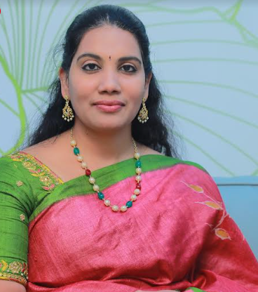
By Dr Chandana Galidevara, Consultant Obstetrician & Gynaecologist, High-risk Pregnancy, Foetal Medicine Specialist & Laparoscopic Surgeon, Ankura Hospital for Women and Children
Bringing a new life into the world is a momentous occasion filled with anticipation and excitement. As expectant parents prepare for the arrival of their little one, one of the most significant decisions they face is choosing between natural birth and Cesarean section (C-section). Both methods of delivery have their own set of considerations, and making an informed decision is paramount for the health and well-being of both the mother and the baby.
In general, the majority of child births worldwide occur through natural vaginal delivery, accounting for approximately 75 to 90⁶% of deliveries. However, the rate of C-sections has been steadily rising globally, reaching around 20 to 25% of all births. Understanding these factors is essential for expectant parents to make an informed decision that aligns with their unique circumstances and preferences.
Medical Considerations
One of the primary factors to consider when choosing between a natural birth and a C-section is the medical condition of both the mother and the baby. Maternal health conditions such as hypertension, diabetes, or heart disease may influence the suitability of vaginal delivery. Similarly, obstetric complications such as Placenta Previa, contracted pelvic, fetal distress, or a breech presentation may necessitate a C-section.
Consulting with healthcare providers, including obstetricians and midwives, is essential. They carefully evaluate the individual’s health condition and determine the safest delivery method for both the mother and the baby, aiming to minimize risks and complications.
Previous Delivery Experience
A woman with a previous childbirth experience can significantly impact her decision regarding the method of delivery. Positive experiences with natural birth may lead to a preference for a vaginal delivery, while traumatic or complicated deliveries may prompt consideration of a C-section. Women who have undergone a C-section in the past may consider vaginal birth after Cesarean (VBAC) after discussion with their healthcare providers and careful assessment of their suitability for VBAC.
It’s essential for expectant parents to reflect on their past experiences and discuss them with their healthcare providers to make an informed decision.
Delivery Process
Understanding the details and implications of the delivery process for both natural birth and C-section is crucial. Natural birth typically involves the onset of labour, progression through different stages of labour, and ultimately, the delivery of the baby through the birth canal. It often results in a shorter recovery time for the mother and promotes early bonding and breastfeeding initiation. On the other hand, a C-section is a surgical procedure that requires anesthesia and involves incisions on the abdomen and uterus to deliver the baby. While C- sections may be necessary in cases of medical emergencies or complications, they generally involve a longer recovery period and a higher risk of infection compared to vaginal delivery.
Personal Preferences and Birth Plans
In the realm of childbirth, personal preferences and birth plans serve as guiding beacons for expectant parents navigating the intricate journey of bringing new life into the world. Some expectant parents may have strong preferences for one method of delivery over the other based on cultural or emotional factors. It’s essential for them to openly communicate with their healthcare providers about their preferences and concerns to ensure that their birth plan aligns with their wishes while prioritizing the health and well-being of both mother and baby. These preferences encompass a spectrum of desires, intentions, and expectations unique to each individual, shaping the trajectory of labour, delivery, and postpartum care.
Support System and Resources
Having a supportive birth team and access to resources such as childbirth education classes and prenatal support services can facilitate the decision-making process. Childbirth education classes provide expectant parents with valuable information about the childbirth process, pain management techniques, and strategies for labour and delivery. Facilities for the provision of epidural, having a supportive partner or support person can provide emotional and practical support throughout labour and childbirth.
Psychological and Emotional Factors
The psychological and emotional readiness of expectant parents is another critical factor to consider when choosing between natural birth and C-section. Pregnancy and childbirth can evoke a range of emotions, including excitement, anxiety, and fear. It’s essential for expectant parents to explore their feelings and seek emotional support from their healthcare providers, family members, or support groups as needed.
In conclusion, choosing between a natural birth and a C-section involves a careful consideration of medical, obstetric, personal, and emotional factors. Expectant parents should work closely with their healthcare providers to weigh the risks and benefits of each delivery method and make an informed decision that aligns with their preferences and circumstances. Addressing issues such as anxiety and alleviating fear helps in making positive choices. By prioritizing safety, well-being, and open communication, expectant parents can embark on their childbirth journey with confidence and peace of mind.
- 0
- By Rabindra

20, Feb 2024
TriLife Hospital Empowers Residents with BLS Training Workshop
Bengaluru, February 20th, 2024 — TriLife Hospital, a leading healthcare provider in Bengaluru, has conducted a specialized Basic Life Saving Skills (BLS) Training Workshop with the Orchidwood Association. The workshop is aimed at equipping residents with essential life-saving skills, focused on handling emergencies involving residents with a focus on children and the elderly. The training program is part of a comprehensive series of workshops to be conducted across apartment complexes in Bengaluru, with the aim of training over 5000 individuals across the city. Led by certified doctors and therapists with extensive experience in emergency response and healthcare, the workshop covered a wide range of topics, including CPR, first response to burn injuries, assessment, and handling of cardiac emergencies, drowning first aid and common first aid training for accidents and mishaps.
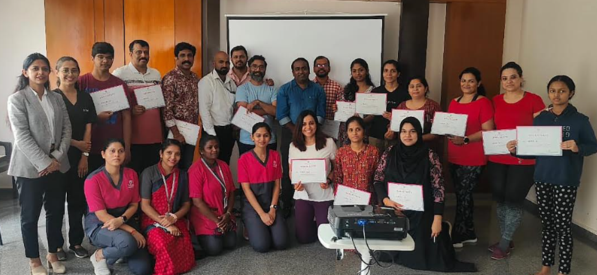
“This BLS training initiative is testament to our commitment to reach as many sections of the public as possible to promote safety and first aid awareness. It provides an indirect effect on improving public health and safety. This initiative aligns with our commitment to educate and empower people with the necessary skills and knowledge on how to respond quickly and effectively in emergency situations. We have already conducted similar programs with the Bengaluru City Police in Hennur and Banaswadi police station limits. As a leading healthcare provider in the city, we remain committed to its mission of saving lives and promoting well-being.” said Dr. Shafiq A M, Director and CEO of TriLife Hospital
TriLife Hospital is in talks with RWA’s across the city and will conduct similar programs with them in future as well .
20, Feb 2024
11-year-old Srilankan Girl Undergoes Rare Deformity Correction using 3D Bone Model Technology at MGM Healthcare
Chennai 20th February 2024: MGM Healthcare, a renowned multi-specialty quaternary care hospital located in the heart of Chennai, proudly announces the successful spine deformity correction surgery performed on an 11-year-old from Sri Lanka. The child, Ms. Shreya (Name Changed), was diagnosed with Neurofibromatosis with L5 S1 Spondyloptosis and Right-sided severe neglected rigid thoracolumbar scoliosis.
The parents of Ms. Shreya had consulted with several doctors at different healthcare facilities in Sri Lanka, but didn’t receive positive outcomes due to high risk involved in surgery. Senior doctors from a private hospital in Sri Lanka recommended the family to seek consultation at MGM Healthcare in Chennai.
Under the guidance of Dr. Karunakaran. S, Director and Senior Consultant, Department of Spine Surgery, MGM Healthcare, and Dr. Vijayaraghavan, Senior Consultant, Department of Spine Surgery, MGM Healthcare, the child underwent a comprehensive evaluation. The doctors advised the family to opt for a deformity correction surgery. Failure of which there is definite risk of progression of deformity and chances of respiratory failure in future leading to risk of life.
Speaking about the procedure Dr. Karunakaran. S, Director and Senior Consultant, Department of Spine Surgery, MGM Healthcare, said “Due to duration of chronic neglection and severity of the deformity the anatomy of spine was completely distorted. Placing screws in such a deformed spine is really challenging hence we advocated 3D bone model technology where a plastic SD bone model will be made similar to patient’s deformed spine pre-operatively based on ct scan. Doctors can plan spine instrumentation before as well as during surgery with the help of 3D model. This creates an avenue to perform surgery more precisely as well as safely.”
The Importance of Early Detection in Pediatric Scoliosis
Scoliosis in children, if left untreated, can lead to severe complications, affecting not only physical health but also emotional well-being. Early detection is critical as it allows for timely intervention and a higher likelihood of successful recovery.
Dr. Vijayaraghavan, Senior Consultant, Department of Spine Surgery, MGM Healthcare, emphasized the significance of early detection, stating, “Ms. Shreya’s case highlights the importance of identifying scoliosis in children at an early stage. Early detection allows for more conservative, less morbid surgical treatment options and significantly improves the chances of a positive outcome.”
Postoperatively, Ms. Shreya exhibited significant clinical improvement. Her successful recovery led to her discharge with tailored advice for ongoing care and rehabilitation.
The successful intervention showcases MGM Healthcare’s commitment to pushing the boundaries of medical expertise and providing world-class care for complex cases.
20, Feb 2024
Prehabilitation – A tool to prevent postoperative complications in patients planned for surgeries
Dr. Swapna Karthikeyan, Tutor, Department of Cardiovascular & Pulmonary Physiotherapy, at Saveetha College of Physiotherapy, Saveetha Institute of Medical and Technical Science, Chennai.
Prehabilitation is a series of interventions carried out in a medical facility prior to a surgical operation. It focuses on the modifiable risk factors, allowing individuals to prepare for treatment by encouraging healthy behaviours and prescribing multimodal programs that include exercise, diet, and psychological therapies depending on their individual needs. In addition, prehabilitation involves assessing medications, encouraging decreased alcohol use, helping patients cease smoking, and optimizing treatment for long-term illnesses including diabetes and anaemia. Prehabilitation aims to increase a patient’s functional capacity before surgery, improve postoperative outcomes, prevent postoperative complications, and assist with the recovery and restoring state following the surgical procedure.
At Saveetha College of Physiotherapy, SIMATS, Chennai, our cardiopulmonary physiotherapy team creates an individualized prehabilitation program based on the patient’s physical, physiological, and psychological state through thorough assessment and management strategies to enhance the efficacy of various treatments, improve the quality of life and functional capacity post-operatively. We counsel the patients regarding the dos and don’ts post-operatively and increase their adherence to a post-operative rehabilitation program. The modern era allows us to use smart devices in planning strategies and monitoring them. This makes it easy and feasible for the patients to take part in the prehabilitation programs.
Prehabilitation helps patients increase their resistance to therapy and enhance their long-term health as it is a component of the rehabilitation continuum.
When deciding on a course of treatment, consider attending a reputable facility like SIMATS, which is renowned for its advanced amenities and can provide individualized treatment tailored to your individual needs.
19, Feb 2024
Chronic Kidney Diseases – Diagnosis & Treatment
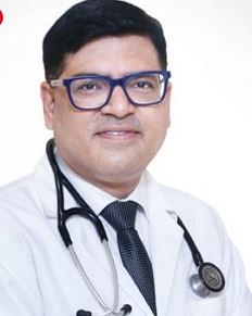
Dr. Vishal Saxena, Senior Director – Nephrology & Renal Transplantation, BLK-MAX Super Speciality Hospital, Delhi
The burden of chronic kidney diseases in India cannot be assessed accurately. The approximate prevalence of CKD is 800 per million population (pmp), and the incidence of end stage renal disease is around 150-200 pmp. Most common cause of kidney disease is diabetes and high blood pressure. Both the diseases are related to our genetics and lifestyle, hence the emphasis is to keep our diabetes and blood pressure under control with medicines, exercise and a blend of healthy food. Blood pressure not only needs to be judiciously controlled , but weather its secondary to kidney disease also needs to be ruled out. Young people developing high bp especially needs to be screened for underlying kidney diseases.often people feel depressed once diagnosed with chronic kidney disease( CKD), hence the whole idea of doctors is now to focus on a holistic approach when dealing with CKD. This involves dietary change, avoiding pain killers , quit smoking and to promptly refer to a nephrologist ,once diagnosed with diabetes , high bp or any form of CKD. Certain kidney diseases are controllable/ treatable hence the need to promptly diagnose the disease and treat it .
Preventive strategies play a very pivotal role in dealing with CKD. Regular health check ups form an integral role in screening of CKD during early phase, which i think should be inculcated in schools, colleges at entry level and while joining a new job or organization. Since treating a CKD patient is more expensive these screening strategies will definitely reduce the financial burden on the patient and on the society as whole. Another most neglected area in our country is lack of physical activity/regular exercise. Regular exercise not only improves physical well being, but also is a stress buster for a lot of patients. All these measures help in creating awareness in coping with the kidney disease and allow the patient to deal with his disease with a positive mindset. Being in regular follow up with one’s kidney physician helps the patient to help discuss strategies to not only control his disease but also to formulate a planned way of dealing with his ailment. India being a big country with patients having varied dietary habits, diet plays a very important role when it comes to kidney diseases, hence patients regular diet counselling is very important. Kidney diseases have a variable presentation; hence a tailor-made therapy is important for each kidney patient.
19, Feb 2024
Spotting STDs Early: Understanding the Telltale Signs
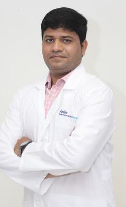
Dr. Basavaraj S Kumbar, Consultant- Internal medicine, Aster Whitefield Hospital, Bengaluru
If you’ve had unprotected sex, it’s important to remember that there’s more at stake than just pregnancy. Not using protection increases the chances of getting sexually transmitted diseases (STDs), which can vary from common infections like chlamydia and gonorrhea to more serious ones like HIV and herpes. It’s essential to know that not all STDs show immediate symptoms. That’s why it’s smart to regularly get tested to catch any surprises early on. Looking after your sexual health means more than just avoiding pregnancy – it’s about protecting yourself and your partner from potential infections.
Sexually transmitted diseases (STDs) are a big worry because they can harm your health in many ways. While some infections cause symptoms like itching, pain, or strange discharges, others don’t show any signs right away. This means you might not even realize you have something wrong inside you. It’s really important to get tested regularly to catch any problems early. The symptoms of these infections can vary a lot, from sores and painful urination to unusual discharge and swollen lymph nodes. If left untreated, STDs can cause serious issues like trouble getting pregnant, damage to your organs, or even cancer. So, it’s not just about you – it’s about taking care of your health and thinking about your future self.
Recognizing the signs
Let’s talk about the common suspects when it comes to sexually transmitted diseases (STDs). These include Bacterial Vaginosis, Chlamydia, Gonorrhoea, Hepatitis, Herpes, HIV, HPV, Mycoplasma Genitalium, Syphilis, and Trichomoniasis. Each of these can bring different symptoms and challenges. For example, Bacterial Vaginosis might give off a fishy odor, while Chlamydia and Gonorrhoea can cause discomfort and unusual discharge. Hepatitis, especially type B, can hide without showing any symptoms, highlighting the importance of vaccination. Herpes shows up with sores, and HIV might initially feel like the flu. HPV, which is quite common, can lead to warts or even cancer, stressing the need for vaccination and regular check-ups. Mycoplasma Genitalium is still being studied in testing, and Syphilis starts with painless sores but can cause serious damage to organs if left untreated. Trichomoniasis typically causes itching, burning, and strange discharge.
Transmission
Exploring how sexually transmitted diseases (STDs) spread and the risks involved, let’s delve into the various transmission routes and their implications. STDs can spread through oral, anal, or vaginal sex, but the level of risk varies for each. While oral sex carries a lower risk for some infections, using condoms and dental dams can help protect against transmission. Anal sex has higher risks because the rectal lining is vulnerable, so it’s crucial to use condoms and lubrication consistently. Vaginal intercourse is a common way for STDs to spread, highlighting the importance of using condoms and getting regular screenings. Also, some infections can spread through skin-to-skin contact, so it’s essential to stay aware even when using condoms.
Is it different for men and women?
Men and women might notice different symptoms when it comes to sexually transmitted diseases (STDs). For example, women may not easily spot genital ulcers, while men might see unusual discharge or sores on their penis. Men usually recognize external symptoms like sores or discomfort, while women might feel pelvic pain or notice subtle changes in vaginal discharge. Genital ulcers might not be as visible in women and could be inside the cervix or vaginal canal. Both men and women can have STDs without showing any symptoms, underlining the need for regular screenings. If left untreated, these infections can lead to problems with fertility. It’s important for partners to communicate openly and use barrier methods consistently to prevent STD transmission.
Treatment and prevention
Treatment and prevention methods for sexually transmitted diseases (STDs) vary depending on the type of infection. Antibiotics can effectively treat bacterial STDs, while viral infections cannot be cured but can be managed with proper medical care. Vaccines against hepatitis A, hepatitis B, and HPV offer an extra layer of protection against certain infections.
Making smart lifestyle choices like abstaining from sex, limiting sexual partners, practicing mutual monogamy, and consistently using condoms significantly lower the risk of getting an STD. Untreated STDs can lead to serious health problems such as infertility, organ damage, illness, cancer, and the spread of other diseases. Routine gynecological check-ups are key for early detection of sexually transmitted diseases (STDs).
19, Feb 2024
Usage of C-ARM technology to improve surgical precision and patient outcomes in orthopedic procedures
Authored by:
Mr. Navjot Singh, Executive Director, Trivitron Healthcare
Usage of C-ARM technology to improve surgical precision and patient outcomes in orthopedic procedures
In the dynamic landscape of modern medicine, technological advancements continue to reshape the way healthcare professionals approach diagnosis and treatment. One such groundbreaking innovation that has significantly transformed orthopedic surgery is the utilization of C-Arm technology. This imaging technology, consisting of a C-shaped arm with an X-ray source and detector, has proven to be instrumental in enhancing surgical precision, real-time visualization, and overall patient outcomes in orthopedic procedures. The C-Arm’s ability to provide high-quality, real-time imaging during surgeries allows orthopedic surgeons to navigate complex anatomical structures with unprecedented accuracy. This capability is particularly valuable in procedures such as joint replacements, spinal surgeries, and trauma interventions. Surgeons can visualize the placement of implants, assess joint alignment, and make real-time adjustments, leading to improved postoperative functionality and reduced complications.
Furthermore, the dynamic nature of C-Arm imaging enables healthcare professionals to confirm the success of interventions immediately, minimizing the need for additional procedures and reducing overall surgical time. As a result, patients experience shorter recovery periods and enhanced overall outcomes. The integration of C-Arm technology into orthopedic practices represents a pivotal step forward, showcasing how innovation continues to revolutionize the field, ultimately benefiting both healthcare providers and the individuals they serve.
Understanding C-Arm Technology:
C-Arm technology refers to a specialized imaging system that consists of a C-shaped arm, an X-ray source, and a detector. This configuration allows for high-quality, real-time imaging during surgical procedures. Originally developed for use in cardiac and vascular procedures, C-Arm technology has seamlessly integrated into orthopedic surgery, providing surgeons with invaluable insights and enhancing their ability to perform complex procedures with unprecedented accuracy.
Benefits of C-Arm Technology in Orthopedic Surgery:
Real-time Imaging and Visualization: One of the primary advantages of C-Arm technology is its ability to provide real-time imaging during surgery. This feature allows orthopedic surgeons to visualize the affected area dynamically, enabling them to make informed decisions and adjustments throughout the procedure. Real-time feedback is particularly crucial in orthopedic surgeries, where precision is paramount.
Improved Surgical Precision: Orthopedic procedures often involve intricate maneuvers, such as joint reconstructions, spine surgeries, and fracture fixations. C-Arm technology facilitates superior precision by offering high-resolution imaging of the targeted anatomy. Surgeons can visualize bone structures, joint spaces, and implant placements with exceptional clarity, leading to more accurate and effective interventions.
Minimized Radiation Exposure: Traditional X-ray imaging methods may expose patients and healthcare professionals to higher levels of radiation. C-Arm technology, however, allows for optimized imaging with lower radiation doses. This reduction in radiation exposure is particularly significant for patients undergoing multiple orthopedic procedures or those who may be more susceptible to the harmful effects of radiation.
Enhanced Workflow and Efficiency: C-Arm technology streamlines the surgical workflow by providing instantaneous images without the need for film development. This efficiency not only saves valuable time during the procedure but also allows for prompt decision-making, ultimately contributing to improved patient outcomes.
Versatility in Applications: The versatility of C-Arm technology extends beyond traditional orthopedic surgeries. It is widely utilized in various interventional procedures, such as pain management, angiography, and orthopedic trauma surgeries. This adaptability makes C-Arm a valuable tool in the hands of healthcare professionals across different specialties.
Educational Opportunities: C-Arm technology facilitates educational initiatives within the medical community. Surgeons can use real-time imaging capabilities to teach and train the next generation of orthopedic specialists. This hands-on approach enhances the learning experience and ensures that future practitioners are well-equipped to leverage the advantages of this innovative technology.
Case Studies and Success Stories:
Numerous case studies and success stories attest to the positive impact of C-Arm technology on orthopedic surgery. For instance, in spine surgeries, where precision is crucial to avoid damage to delicate structures, C-arm imaging has significantly reduced the risk of complications. Similarly, in joint replacement surgeries, accurate placement of implants is essential for long-term success, and C-Arm technology has proven instrumental in achieving optimal alignment and stability.
Future Directions and Challenges:
As C-Arm technology continues to evolve, ongoing research aims to address challenges and further enhance its capabilities. The integration of artificial intelligence and machine learning algorithms into C-Arm systems holds promise for automated image interpretation and enhanced surgical guidance. However, challenges such as cost considerations, accessibility, and training for healthcare professionals need to be addressed to ensure widespread adoption and utilization of this technology.
In the end, it can be concluded that the integration of C-Arm technology into orthopedic surgery represents a milestone in the pursuit of improved patient outcomes and surgical precision. The real-time imaging capabilities, enhanced workflow, and versatility of applications make C-Arm technology an invaluable tool for orthopedic surgeons. As technology continues to advance, it is poised to play a pivotal role in shaping the future of orthopedic surgery, setting new standards for precision, efficiency, and overall excellence in patient care. C-Arm technology stands as a testament to the ongoing commitment of the medical community to embrace innovation and elevate the standards of healthcare delivery.
19, Feb 2024
Supporting the Diagnosis of Bone-related Diseases, FUJIFILM India Installs an Advanced DEXA Machine in Delhi
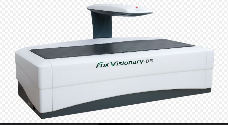
New Delhi, 19 February 2024: FUJIFILM India, a pioneer in diagnostic imaging solutions, has announced the installation of its powerful Dual-Energy X-ray Absorptiometry (DEXA) machine “FDX Visionary-DR” at the grand inauguration of the Center for Sports Injury in Delhi. The event took place on the 11th of February with the honourable dignitaries from centre alongside the presence of top sporting icons. The Center for Sports Injury is the premier sports health and wellness facility that specializes in Orthopedics, Sports Surgery, Sports Medicine, Sports Imaging, Sports Physiotherapy, Rehabilitation, Fitness and Counseling. The initiative aims to deliver high-quality and fast bone-density scans, commonly known as DEXA scans, to diagnose bone-related health conditions or evaluate the risk of developing such problems. With its innovative machinery, FUJIFILM India endeavors to make excellent diagnostic capabilities accessible to bone densitometry practitioners nationwide.

Bone-related health diseases such as osteoporosis are widespread in India, with estimates indicating that 61 million individuals suffer from this condition. Furthermore, the incidence of osteoporosis tends to occur 10 – 20 years earlier here, in comparison to Western countries. Tackling this, the bone densitometry scan serves as an effective tool to accurately predict the chances of fractures or abnormalities in the upcoming years. FUJIFILM India’s FDX Visionary-DR machine delivers a complete diagnostic experience with a wide range of applications and routine examinations for Orthopedics, Pediatrics, Lateral Spine, Morphometry and Whole Body. It features a breakthrough 3D-DXA technology that uses routine bone-mineral-density images to create 3D models of the femur, bringing in new information about bone structure for accurate diagnosis and adapted treatment procedures. Based on a 4-line, 64-element multi-array detector, the machine’s remarkable 2D Fan Beam is designed to provide the highest image resolution for a precise diagnosis and perform quick 15-second assessments per site, making it one of the most powerful solutions available in the market. Additionally, it offers advanced fat, muscle and lean medical parameters through its BODY COMPOSITION tool which is essential in sports medicine, weight management and medical practice fields.
“In India, the growing prevalence of bone-related health conditions highlights the urgent need for preventive measures in healthcare. As a leader in diagnostic imaging solutions, FUJIFILM India recognizes the critical role of early detection and timely intervention in mitigating the impact of bone-related diseases on patients and society as a whole. Thus, we are proud to introduce this FDX Visionary-DR machine in Delhi as it possesses the potential to revolutionize the bone health-related diagnostic imaging segment. Through breakthrough imaging technologies, strategic partnerships and awareness drives, our mission is to effectively diagnose, treat and manage bone health conditions and pave the way towards a healthier future for India” said, Mr. Chander Shekhar Sibal, Vice President & HOD, Healthcare Business, FUJIFILM India.
Osteoporosis occurs due to excessive bone loss, insufficient bone formation or a combination of both factors. Consequently, bones become weak, making them susceptible to fractures even from minor accidents or from everyday actions like sneezing or bumps in severe cases. Addressing this, dual-energy X-ray absorptiometry or DEXA scans measure bone loss by utilizing a minimal dose of ionizing radiation to generate images of internal body structures such as the hip, spine and forearm. It also helps with the periodic monitoring of body fitness parameters like densitometry and body composition analysis, examining the density of peripheral central body parts as well as visceral and subcutaneous fat analysis.
Dr Pushpinder – an eminent figure with over 30 years of Experience in the field of Sports Medicine, Joint Replacement Surgery & Arthroscopic Surgery (KEYHOLE SURGERY) nationally & internationally while embracing the meaningful installation said, “The alarming estimate of millions suffering from bone-related conditions like osteoporosis highlights a pressing issue which is affecting the quality of life across the nation. Also, the fact that these critical diseases tend to occur earlier in India, necessitates urgent attention to preventive care and supporting action to improve the situation. So, we take great pride in welcoming this sophisticated DEXA machine, courtesy of FUJIFILM India, at our facility to deliver reliable diagnostic services in Delhi and help communities lead a healthier life.”
FUJIFILM India reaffirms its commitment to reshaping India’s healthcare sector with the exceptional FDX Visionary-DR machine, equipping medical professionals with future-ready diagnostic technologies to provide crucial and urgent patient care.
19, Feb 2024
Pediatric Neurologist , Dr. Vykunta Raju K. N., Receives Prestigious Award for Outstanding Contribution to Child Neurology
Bengaluru, February 19, 2024: In a momentous occasion, Dr. Vykunta Raju K. N., Professor and Head of the Department at Indira Gandhi Institute of Child Health, has been honored with the Outstanding Contribution Award for Child Neurology by the Association of Child Neurology, India (AOCN). Dr. Raju, with over 15 years of dedicated service in pediatric neurology, has played a pivotal role in the diagnosis and management of rare and neurological disorders.
The accolade comes as recognition for Dr. Vykunta Raju’s tireless efforts in advancing the understanding and treatment of rare diseases, particularly in pediatric neurology. Despite the challenges faced in the field, he remains committed to improving the lives of children and families affected by these disorders. Dr. Raju has published over 200 publications focusing on original reviews and case reports on rare disorders to enhance awareness, understanding prevalence, and studying epidemiological factors in the Indian population.
While expressing his happiness for the award recognition, Dr Raju, said, “I am feeling happy to receive this award. I have been working in pediatric neurology for the past 15 years, and we have done a lot of work. Though the recognition has come a little late, I still feel very happy on getting recognized for the work I have contributed. In pediatric neurology, you will be managing a lot of rare disorders, most of the time. In earlier days, these rare diseases were neglected and treated as cerebral palsy. But now, with improvement in genetics, we are able to diagnose these disorders. Arriving at the right diagnosis was a huge challenge in the earlier days. Compared to that, diagnosis is easier, but getting proper treatment is still an issue. Treatment is not available for many disorders; and even if it is available, the cost of the medicines is very high. So, most of our patients are unable to afford these treatments.”
Speaking on Rare Disease Awareness Month (February), Dr. Raju highlighted the importance of accurate diagnosis and the need for FDA-approved treatments, addressing the difficulties faced by patients, particularly in accessing expensive medications for some disorders like Spinal Muscular Atrophy. In most of the countries, these medications are made available mostly by Government funding. So, if it’s made available in India also, it will benefit both the children and their families.
19, Feb 2024
Amblyopia – if Untreated, It Can Affect a Child’s Vision Permanently
Amblyopia is the most common vision issue that affects kids. Amblyopia (lazy eye) causes blurry vision in one eye when something affects how a child’s eyes are developing. As their brain ignores the weaker eye, that eye drifts out of position. It’s rare, but amblyopia can affect both eyes at the same time. It affects around 5% of children younger than 15 years. Eye care specialist will treat amblyopia by making child’s brain use their weaker eye to see.
This will repair and strengthen the connection between your child’s brain and both their eyes to correct the amblyopia. Most kids need amblyopic treatment at least for few months. Treatment can be done by eye patching, atropine therapy or by spectacle correction. This can include targeted education, awareness campaigns, and policy interventions.
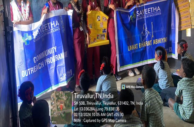
As a part of UBA and community outreach program, students and faculty of “Saveetha College of Allied Health Sciences” reached kondancherry village, Tiruvallur District, Tamil Nadu and conducted an awareness program by stating the Importance of eye screening in children to prevent amblyopia.