


25, Mar 2026
End of a Long Wait: Harish Rana Passes Away After 13 Year Coma
Harish Rana, known as one of the first individuals in India to be permitted passive euthanasia, passed away on Tuesday at the All India Institute of Medical Sciences (AIIMS), bringing an end to a long and difficult chapter that spanned over 13 years.
Rana, 31, had been in a coma since 2013 following a severe medical condition that left him unresponsive. For more than a decade, his life was sustained through continuous medical care, largely managed at his home in Ghaziabad by family members who remained by his side through the prolonged ordeal.
Earlier this month, on March 14, he was shifted to the palliative care unit of Dr. B.R. Ambedkar Institute Rotary Cancer Hospital at AIIMS-Delhi as his condition required specialized end-of-life support. Medical teams provided comfort care during his final days.
Rana’s case had drawn attention for its legal and ethical dimensions, particularly around the issue of passive euthanasia in India. It highlighted the emotional and financial strain faced by families caring for patients in prolonged vegetative states, while also bringing focus to the need for compassionate end-of-life care policies.
Despite the legal and medical complexities surrounding his condition, at the heart of the story was a family’s unwavering commitment. For years, his loved ones continued to hope, care, and advocate, navigating both personal grief and public attention.
His passing marks not just the end of a life, but the conclusion of a deeply human story—one that sparked conversations about dignity, medical ethics, and the realities of long-term critical care.
Harish Rana is remembered not only for the circumstances of his case but also for the quiet resilience shown by those who stood beside him throughout his journey.

- 0
- By Neel Achary

25, Mar 2026
India Unveils Multi-Centre Study on Ayurveda Support in TB Care
New Delhi, Mar 25 (BNP): The Ministry of Ayush, in collaboration with the Department of Biotechnology (DBT), has announced a joint clinical study to evaluate Ayurveda as an adjunct therapy in tuberculosis (TB) treatment, marking a step toward integrative healthcare in India.
The announcement was made on World Tuberculosis Day at a high-level event held at Vigyan Bhawan, attended by senior policymakers, scientists, and healthcare experts.
Union Minister of State (Independent Charge) for Science and Technology, Dr. Jitendra Singh, said India is making steady progress in its fight against TB through science-driven innovation and integrated healthcare approaches.
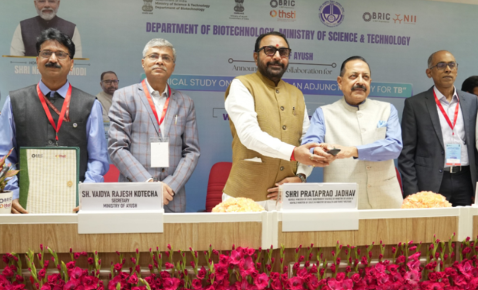
“The collaborative clinical study reflects our commitment to evidence-based innovation, combining modern biomedical research with traditional knowledge systems,” he said, adding that efforts are also focused on addressing challenges such as drug resistance and undernutrition.
Minister of State (Independent Charge) for Ayush, Prataprao Jadhav, emphasised that TB treatment must go beyond eliminating infection to ensuring complete patient recovery and improved quality of life.
“With this approach, Ayurveda and other Ayush systems can play a supportive role in enhancing recovery, nutrition, and overall well-being,” he said.
The study, titled “Clinical Study on Ayurveda as an Adjunct Therapy for Tuberculosis”, will be conducted as a multi-centre trial across premier institutions, including AIIMS, JIPMER, and NEIGRIHMS.
Led by the Central Council for Research in Ayurvedic Sciences (CCRAS) in partnership with DBT, the 24-month study aims to assess the efficacy, safety, and tolerability of an Ayurveda regimen alongside standard anti-tuberculosis treatment (ATT), combined with nutritional support.
Officials said the initiative is expected to generate scientific evidence on improving recovery rates, nutritional outcomes, and quality of life among TB patients.
The programme also witnessed the exchange of a memorandum of understanding between BRIC-Translational Health Science and Technology Institute (THSTI) and CCRAS to formalise institutional collaboration.
The government reaffirmed its commitment to advancing evidence-based integrative healthcare solutions to tackle public health challenges and accelerate progress toward TB elimination.
22, Mar 2026
IHCL Hotels in Tamil Nadu Reinforce Responsible Hospitality, Mark Four Years of Paathya with Bharat Eksaath Walkathon
CHENNAI, MARCH 22, 2026: Indian Hotels Company (IHCL), India’s largest hospitality company, continues to advance its sustainability and social impact agenda through Paathya, its ESG+ framework.
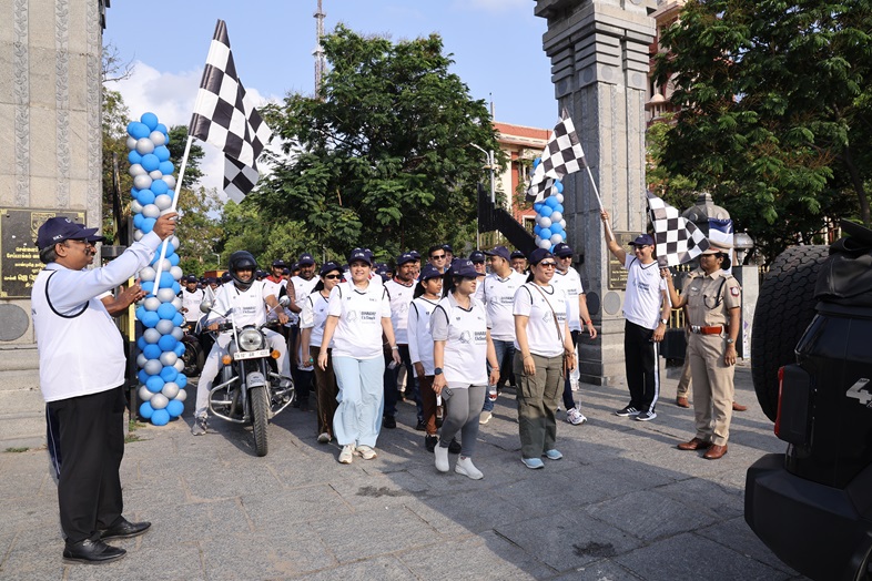
Photo caption: Dr. S. Armstrong, Vice-Chancellor Convener Committee, University of Madras, Megalina Iden, DC Traffic – East, and Ian Dubier, Area Director Operations for Tamil Nadu and General Manager of Taj Coromandel, flagging off IHCL’s Bharat EkSaath Walkathon at University of Madras in Chennai.
Marking its fourth year, IHCL reinforced its commitment to responsible tourism with Bharat EkSaath Walkathon – Tamil Nadu edition, an inclusive 3 km walk that brought together associates, their families, local partners, vendors and community members. This was held simultaneously across 14 countries in over 200 locations. The funds raised through the walkathon support the Taj Public Service Welfare Trust (TPSWT), reinforcing IHCL’s approach to social impact.
Teams from Taj Coromandel, Taj Connemara, Taj Club House, Taj Fisherman’s Cove Resort & Spa Chennai, Taj Wellington Mews Chennai, Vivanta Chennai IT Expressway, Ginger Chennai, OMR, Ginger Chennai, Vadapalani, Ginger Chennai, IITM, TajSATS, Vivanta Coimbatore, Savoy, Ooty – IHCL SeleQtions, Gateway Madurai and Gateway Coonoor participated in the 3-km walk.
The Walkathon commenced from the University of Madras and was flagged off by distinguished guests, Dr. S. Armstrong Vice-Chancellor Convener Committee, University of Madras, and Megalina Iden, DC Traffic – East. The route began at the University of Madras, proceeded along Madras Beach Road, and concluded back at the University campus. The event concluded with a hearty breakfast, followed by the distribution of medals and certificates to all participants.
21, Mar 2026
Ayushman Bharat: From Health Access to Health Security in India
For decades, India’s healthcare system faced a difficult balancing act—expanding access while keeping care affordable. Millions of families, especially in rural and low-income communities, often delayed or avoided treatment due to high costs. A single hospitalization could push households into debt, turning a health crisis into a financial one.
The launch of Ayushman Bharat in 2018 marked a turning point in this journey. More than just a government scheme, it introduced a new vision—one where healthcare is not limited to access, but extends to security, dignity, and long-term well-being.
From Health Access to Health Security: Ayushman Bharat is reshaping Indian public healthcarehttps://t.co/byAyCzSSVf
via NaMo App pic.twitter.com/1FHPwT0tvr
— PMO India (@PMOIndia) March 20, 2026
A Transformative Vision for Public Health
Ayushman Bharat reimagines healthcare as a continuous system rather than a one-time service. It connects prevention, treatment, infrastructure, and digital innovation into a unified framework. This integrated approach ensures that individuals are not only treated when they fall ill but are also supported in staying healthy.
Today, it stands as one of the largest publicly funded healthcare initiatives in the world, reflecting India’s commitment to inclusive and equitable healthcare.
The Four Pillars of Ayushman Bharat
At the heart of this transformation are four key pillars that work together to deliver comprehensive care:
1. Ayushman Arogya Mandirs: Bringing Care Closer to Communities
Formerly known as Health and Wellness Centres, Ayushman Arogya Mandirs have reshaped primary healthcare in India. Spread across villages and towns, these centers provide essential services right at the community level.
They focus on:
- Maternal and child healthcare
- Immunization and nutrition
- Screening for non-communicable diseases such as diabetes, hypertension, and cancers
- Basic medicines and diagnostic services
- Wellness activities, including yoga
By prioritizing early detection and prevention, these centres reduce the need for costly treatments later.
2. Pradhan Mantri Jan Arogya Yojana (PM-JAY): Financial Protection for Millions
The insurance component of Ayushman Bharat ensures that healthcare is affordable. It provides cashless treatment coverage of up to ₹5 lakh per family per year for secondary and tertiary hospitalization.
This has been especially significant for economically vulnerable families, who previously had limited access to quality healthcare. Today, they can seek treatment in empaneled hospitals without worrying about immediate financial burden.
3. Building Stronger Systems with PM-ABHIM
The Pradhan Mantri Ayushman Bharat Health Infrastructure Mission focuses on strengthening the healthcare system itself.
Investments under this mission include:
- Development of critical care blocks in districts
- Expansion of diagnostic and laboratory networks
- Strengthening disease surveillance systems
- Improving emergency response capabilities
This pillar ensures that healthcare delivery is not only accessible but also reliable and prepared for future challenges.
4. Digital Health Revolution
Technology is playing a key role in making healthcare more efficient and transparent. The Ayushman Bharat Digital Mission aims to create a seamless digital ecosystem.
Through digital health IDs and integrated records:
- Patients can access their medical history anywhere
- Doctors can make better-informed decisions
- Services become faster and more coordinated
Telemedicine platforms like eSanjeevani have further expanded access, especially in remote areas, enabling millions of consultations without the need for travel.
Measurable Impact: Changing Lives at Scale
The scale of Ayushman Bharat is matched by its real-world impact:
- Financial Relief: Families are saving substantial amounts on medical expenses, reducing dependence on loans or selling assets.
- Wider Coverage: Tens of crores of citizens have been enrolled, including a major expansion to cover senior citizens above 70 years.
- Stronger Public Hospitals: Government hospitals are being revitalized through better funding and increased patient flow.
- Preventive Healthcare: Large-scale screenings are helping detect diseases early, improving health outcomes and reducing long-term costs.
This shift towards prevention marks a significant evolution in India’s healthcare approach.
From Treatment to Prevention
One of the most important changes brought by Ayushman Bharat is the growing focus on preventive care. Instead of reacting to illness, the system now emphasizes early diagnosis and regular health monitoring.
Screenings for conditions like diabetes, hypertension, and cancer are becoming more common at the community level. This not only improves quality of life but also reduces the overall burden on hospitals.
Strengthening Healthcare for the Future
Beyond immediate benefits, Ayushman Bharat is building long-term resilience in India’s healthcare system. New infrastructure, improved digital connectivity, and community participation are creating a foundation for sustainable growth.
The government’s continued investment and policy support indicate a strong commitment to expanding and improving the scheme in the coming years.
Challenges and the Road Ahead
While the progress is significant, challenges remain. Ensuring consistent quality across regions, addressing gaps in rural infrastructure, and increasing awareness about available services are ongoing priorities.
However, the direction is clear—towards a system that is inclusive, efficient, and centered on people’s needs.
Conclusion: A Healthier, More Secure India
Ayushman Bharat represents a bold step in India’s development journey. It moves beyond the idea of healthcare as a service and embraces it as a right and a safeguard for every citizen.
By combining financial protection, accessible services, strong infrastructure, and digital innovation, it is creating a healthcare system that is not only larger but also better.
As India continues to evolve, Ayushman Bharat stands as a powerful example of how thoughtful policy and collective effort can transform lives—ensuring that health is not a privilege, but a promise fulfilled for all.
21, Mar 2026
Centre Holds ‘Suraksha Sankalp’ Workshop to Strengthen HIV Response in Delhi, Haryana
New Delhi, March 21 (BNP): The Ministry of Health and Family Welfare, through the National AIDS Control Organisation (NACO), convened the ‘Suraksha Sankalp Karyashala’ in Delhi to accelerate district-level HIV/AIDS response, with a focused approach on Haryana and the national capital.
The workshop, chaired by Rakesh Gupta, aimed to strengthen data-driven, district-specific strategies to improve prevention, testing, treatment, and overall service delivery for HIV/AIDS.
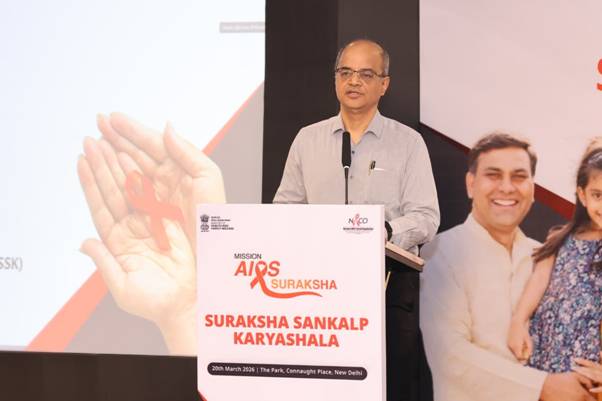
In his keynote address, Dr. Gupta stressed that HIV/AIDS continues to be a significant public health challenge, requiring coordinated and sustained efforts across all levels of governance. He highlighted the global 95:95:95 targets, which aim to ensure that 95 per cent of people living with HIV know their status, 95 per cent of those diagnosed receive treatment, and 95 per cent of those on treatment achieve viral suppression.
Reviewing current progress, officials noted that Delhi faces critical gaps, with only about 70 per cent of identified individuals linked to treatment, while Haryana has achieved an encouraging cascade of approximately 81:83:95, though further improvements are needed in diagnosis and treatment linkage.
The workshop also emphasised the elimination of mother-to-child transmission of HIV, which can be prevented through timely testing, counselling, and treatment during pregnancy, childbirth, and breastfeeding.
Officials said 219 districts across the country have been identified as priority areas for intensified HIV interventions, including 7 districts in Delhi and 11 in Haryana. District-level teams participated in the workshop to present progress, identify challenges, and develop targeted action plans.
Dr. Gupta called for a “whole-of-system” approach, urging stronger coordination between national, state, and district stakeholders to address gaps in awareness, testing, and treatment adherence.
He also outlined India’s goal of bringing HIV/AIDS under control by World AIDS Day 2027, with an enhanced target of achieving 95:95:99 metrics in the coming years.
The workshop forms part of India’s broader strategy to end AIDS as a public health threat by 2030, focusing on evidence-based interventions, improved treatment access, and reducing stigma and discrimination.
21, Mar 2026
Centre Sets Up National Dental Commission, Replaces Dental Council of India
New Delhi, March 21 (BNP): The Government of India has constituted the National Dental Commission (NDC), marking a major reform in dental education and regulation, with the aim of improving quality and aligning standards with global benchmarks.
The new framework, notified on March 19, 2026, replaces the Dental Council of India and comes into effect immediately. With this, the Dentists Act, 1948 has been repealed, and the existing regulatory structure has been dissolved.
Officials said the move represents a shift towards a transparent, accountable, and quality-driven regulatory system, replacing the earlier elected body with a more streamlined governance model.

The NDC will function through three autonomous boards to oversee key areas. The Undergraduate and Postgraduate Dental Education Board will regulate academic standards, while the Dental Assessment and Rating Board will handle accreditation and institutional evaluation. The Ethics and Dental Registration Board will be responsible for professional conduct and registration of practitioners.
Dr. Sanjay Tewari has been appointed as Chairperson of the Commission, with Dr. Mousumi Goswami as a part-time member.
Other key appointments include Dr. Chandrashekhar Janakiram as President of the Education Board, Lt. Gen. (Retd.) Nanda Kishore Sahoo heading the Assessment and Rating Board, and senior professionals appointed across all boards to ensure effective implementation.
The Commission will be responsible for framing regulations, assessing and rating dental institutions, promoting research, evaluating human resource requirements, and setting standards for education, community dental care, and professional ethics.
It will also frame guidelines for fee regulation in private dental colleges, a move aimed at improving transparency and affordability in dental education.
Officials said the reform is expected to enhance the quality of dental education, strengthen healthcare delivery, and expand access to affordable oral healthcare services across the country.
20, Mar 2026
Large imaging study changes understanding of the origins of Parkinson’s rest tremor
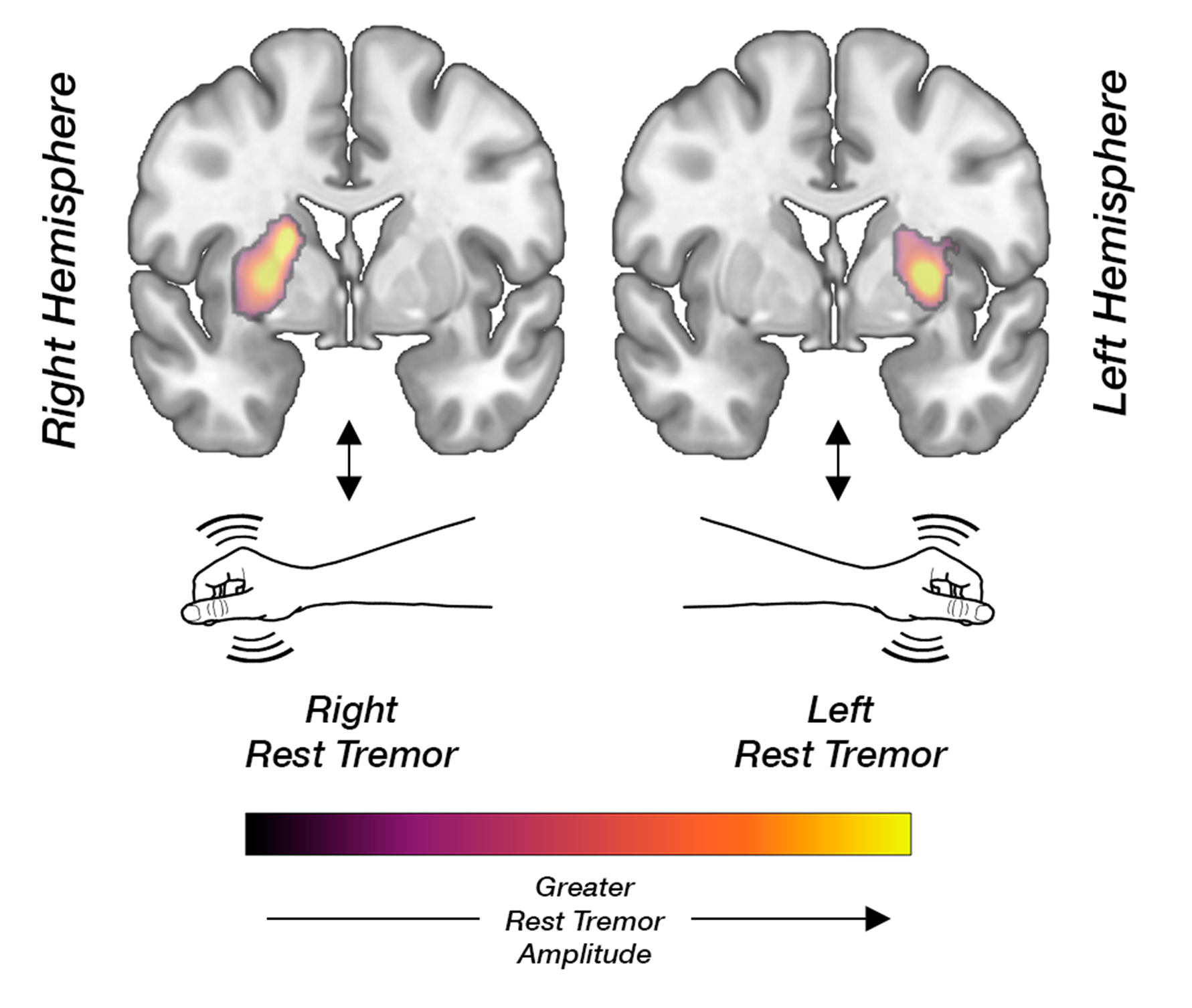
| A Finnish clinical imaging study shows that rest tremor in Parkinson’s disease is not explained by greater dopamine loss. In contrast, tremor appears to be associated with relatively better-preserved dopamine function. |
|
|
|
Researchers from the University of Turku and Turku University Hospital, Finland, analysed clinical data and dopamine transporter (DAT) imaging data from 414 Finnish patients. The cohort consisted of patients examined in routine clinical practice for uncertain parkinsonism or tremor, making the findings exceptionally well generalisable to real-world clinical settings. The results were published on 19 March 2026 in Neurology®, the prestigious medical journal of the American Academy of Neurology. The cardinal motor symptoms of Parkinson’s disease are slowness of movement (bradykinesia), muscle stiffness (rigidity), and rest tremor. Bradykinesia and rigidity are known to reflect degeneration of dopamine-producing neurons. Because most brain pathways cross, this association is typically observed in the striatum on the side opposite to the symptoms. In contrast, the biological basis of rest tremor has long remained uncertain. The study revealed a clear and consistent phenomenon: rest tremor was associated with higher dopamine transporter binding in the striatum on the same side as the tremor. Other cardinal motor symptoms, however, showed the expected correlation with dopamine deficits in the opposite hemisphere. “These results show that more severe rest tremor is not simply a marker of more advanced damage to the dopamine system,” says the lead author, Neurologist Kalle Niemi, MD, PhD. “Tremor appears to involve a partly distinct neurobiological mechanism.” The findings confirm the group’s earlier observations made using data from the international Parkinson’s Progression Markers Initiative (PPMI) cohort, where a novel imaging analysis technique developed by the research team was first applied. The replication of the results in an independent and clinically representative cohort strengthens the reliability of the observed phenomenon. “Our findings support the view that different symptoms of Parkinson’s disease may be driven by partly distinct neural network and neurotransmitter mechanisms,” Niemi explains. “This may help explain why tremor behaves differently from symptoms such as bradykinesia.” Using the same methodological framework, the research team also demonstrated that key non-motor symptoms of Parkinson’s disease including depression, anxiety, and REM sleep behaviour disorder are primarily linked to monoaminergic systems other than dopamine. Taken together, these findings reinforce the concept of Parkinson’s disease as a complex brain disorder involving alterations across multiple neural networks and neurotransmitter systems. A more precise understanding of the biological differences between symptoms may, in the future, enable the development of more targeted and personalised treatment approaches. |
20, Mar 2026
Kaida BioPharma Announces Executive Leadership Team to Advance KAD-101, Built for the Ovarian Cancer Gap
FORT LAUDERDALE, Fla., Mar 20 – Kaida BioPharma, a biotechnology company developing targeted therapies for women with hard-to-treat gynecologic cancers, announced its executive leadership team and board. Each member brings decades of experience in biotechnology, clinical development, regulatory strategy, manufacturing, finance, and commercialization to help advance Kaida’s lead product candidate, KAD-101, toward first-in-human clinical evaluation. The company is developing KAD-101 for recurrent ovarian cancer, where treatment options narrow quickly and durable control remains limited.
Kaida BioPharma is building KAD-101, a next-generation prolactin receptor antagonist for the ovarian cancer gap, the space where patients are too often left cycling through treatment with too few durable options.
In addition to her role as Chairperson of the Board, Kaida founder Dr. Stella Vnook has been named Acting Chief Executive Officer. Dr. Vnook is an accomplished biotechnology founder, executive, and advisor with extensive experience guiding early-stage therapeutics from scientific foundation through capital formation. At Kaida, she leads corporate strategy, development planning, and investor engagement.
“The science brought me here, and the women out there still fighting are why I stay. At Kaida, we are building for the ovarian cancer gap, for patients whose options narrow too fast and whose disease still outpaces available therapies,” said Dr. Vnook. “By targeting the survival signals that keep these tumors alive, we believe KAD-101 has the potential to help overcome treatment resistance and create a new path toward more durable control. More options mean more time, and more time means everything.”
Craig Pierson, a founder alongside Dr. Vnook, has been appointed Director at Kaida BioPharma. Mr. Pierson is a life sciences investor and company builder with more than 25 years of experience in investment banking and private capital formation. He provides strategic oversight and financing expertise.
George E. Peoples, MD, FACS has been appointed fractional Chief Medical Officer. Dr. Peoples is a surgical oncologist with decades of experience in translational oncology and clinical development. He is also the founder of the Cancer Vaccine Development Program, Cancer Insight, and LumaBridge. At Kaida, he helps guide clinical strategy and IND planning.
John Langenheim, PhD has been appointed fractional Chief Scientific Officer. Dr. Langenheim is a molecular biologist with more than 20 years of experience in therapeutic protein design and preclinical development. His scientific leadership has been instrumental in advancing KAD-101 toward clinical evaluation.
Pamela Swiggard has been appointed Head of Regulatory Affairs. She is a pharmaceutical executive with prior experience at companies including Pfizer and Endo Pharmaceuticals. She brings deep expertise across the product lifecycle, from development through regulatory approval and commercialization. At Kaida, she leads regulatory strategy and FDA engagement in support of IND readiness for KAD-101.
Eric Hacherl, PhD has been appointed Head of Manufacturing. Dr. Hacherl is a senior pharmaceutical operations leader with more than 25 years of experience in biologics manufacturing, cGMP compliance, and process development. At Kaida, he is responsible for helping ensure clinical supply readiness for KAD-101.
Mark Booth has been appointed Chief Commercial Officer. Mr. Booth is a seasoned pharmaceutical and biotechnology executive with more than 25 years of experience building commercial strategies for oncology and rare disease products. His background includes leadership across launch planning, brand strategy, access, and market development for important oncology products, including EMEND, EMEND IV, Zolinza, vintafolide, KEYTRUDA, and the avutometinib plus defactinib combination for low grade serous ovarian cancer. At Kaida, he is responsible for commercial strategy for KAD-101, including positioning, market development, and long-term access planning.
20, Mar 2026
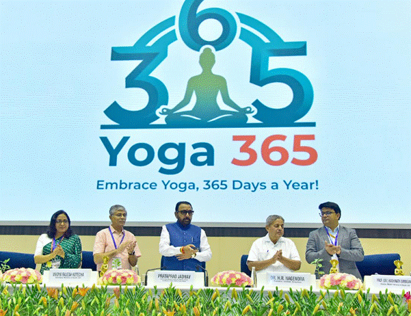
Government Launches ‘Yoga 365’ Campaign to Make Daily Yoga a National Habit
NEW DELHI, March 20: The Government has launched Yoga 365, a nationwide initiative aimed at transforming yoga from an annual observance into a daily practice, with a focus on integrating wellness into everyday life.
The campaign was unveiled during the launch of Yoga Mahotsav–2026 at Vigyan Bhawan, marking the 100-day countdown to the 2026 edition of the International Day of Yoga.
Prataprao Jadhav said the initiative seeks to extend the spirit of the annual yoga day into a sustained, year-round movement. “The aim is to make yoga an integral part of citizens’ daily lives,” he noted.
As part of the campaign, the Morarji Desai National Institute of Yoga signed a memorandum of understanding with wellness platform Habuild to offer free daily online yoga sessions, enabling wider accessibility across regions and age groups.
Officials highlighted that while awareness of traditional wellness systems is high—over 95 percent in both rural and urban India, according to the National Sample Survey—regular practice remains significantly lower. The initiative aims to bridge this gap by encouraging behavioural change and routine adoption.
The programme will focus on community outreach, institutional partnerships, and digital engagement, promoting yoga across schools, workplaces, and neighbourhood groups.
Vaidya Rajesh Kotecha said Yoga 365, along with new therapeutic yoga protocols, is designed to make yoga more practical and relevant for daily health needs.
Describing it as a step towards building a culture of preventive healthcare, Monalisa Dash said the campaign aims to ensure yoga becomes a sustainable lifestyle practice across households.
The initiative builds on the growing global reach of the International Day of Yoga, which has evolved into a major wellness movement since its inception in 2015. Officials said the next phase will focus on embedding yoga into everyday routines, beyond symbolic celebrations.
With the countdown to IDY 2026 underway, the government expressed confidence that Yoga 365 could help convert widespread awareness into consistent daily practice, strengthening both individual health and collective well-being.
19, Mar 2026
Cathy Tie Launches Origin Genomics to Advance Responsible Germline Gene Correction in the United States