


19, Feb 2024
Usage of C-ARM technology to improve surgical precision and patient outcomes in orthopedic procedures
Authored by:
Mr. Navjot Singh, Executive Director, Trivitron Healthcare
Usage of C-ARM technology to improve surgical precision and patient outcomes in orthopedic procedures
In the dynamic landscape of modern medicine, technological advancements continue to reshape the way healthcare professionals approach diagnosis and treatment. One such groundbreaking innovation that has significantly transformed orthopedic surgery is the utilization of C-Arm technology. This imaging technology, consisting of a C-shaped arm with an X-ray source and detector, has proven to be instrumental in enhancing surgical precision, real-time visualization, and overall patient outcomes in orthopedic procedures. The C-Arm’s ability to provide high-quality, real-time imaging during surgeries allows orthopedic surgeons to navigate complex anatomical structures with unprecedented accuracy. This capability is particularly valuable in procedures such as joint replacements, spinal surgeries, and trauma interventions. Surgeons can visualize the placement of implants, assess joint alignment, and make real-time adjustments, leading to improved postoperative functionality and reduced complications.
Furthermore, the dynamic nature of C-Arm imaging enables healthcare professionals to confirm the success of interventions immediately, minimizing the need for additional procedures and reducing overall surgical time. As a result, patients experience shorter recovery periods and enhanced overall outcomes. The integration of C-Arm technology into orthopedic practices represents a pivotal step forward, showcasing how innovation continues to revolutionize the field, ultimately benefiting both healthcare providers and the individuals they serve.
Understanding C-Arm Technology:
C-Arm technology refers to a specialized imaging system that consists of a C-shaped arm, an X-ray source, and a detector. This configuration allows for high-quality, real-time imaging during surgical procedures. Originally developed for use in cardiac and vascular procedures, C-Arm technology has seamlessly integrated into orthopedic surgery, providing surgeons with invaluable insights and enhancing their ability to perform complex procedures with unprecedented accuracy.
Benefits of C-Arm Technology in Orthopedic Surgery:
Real-time Imaging and Visualization: One of the primary advantages of C-Arm technology is its ability to provide real-time imaging during surgery. This feature allows orthopedic surgeons to visualize the affected area dynamically, enabling them to make informed decisions and adjustments throughout the procedure. Real-time feedback is particularly crucial in orthopedic surgeries, where precision is paramount.
Improved Surgical Precision: Orthopedic procedures often involve intricate maneuvers, such as joint reconstructions, spine surgeries, and fracture fixations. C-Arm technology facilitates superior precision by offering high-resolution imaging of the targeted anatomy. Surgeons can visualize bone structures, joint spaces, and implant placements with exceptional clarity, leading to more accurate and effective interventions.
Minimized Radiation Exposure: Traditional X-ray imaging methods may expose patients and healthcare professionals to higher levels of radiation. C-Arm technology, however, allows for optimized imaging with lower radiation doses. This reduction in radiation exposure is particularly significant for patients undergoing multiple orthopedic procedures or those who may be more susceptible to the harmful effects of radiation.
Enhanced Workflow and Efficiency: C-Arm technology streamlines the surgical workflow by providing instantaneous images without the need for film development. This efficiency not only saves valuable time during the procedure but also allows for prompt decision-making, ultimately contributing to improved patient outcomes.
Versatility in Applications: The versatility of C-Arm technology extends beyond traditional orthopedic surgeries. It is widely utilized in various interventional procedures, such as pain management, angiography, and orthopedic trauma surgeries. This adaptability makes C-Arm a valuable tool in the hands of healthcare professionals across different specialties.
Educational Opportunities: C-Arm technology facilitates educational initiatives within the medical community. Surgeons can use real-time imaging capabilities to teach and train the next generation of orthopedic specialists. This hands-on approach enhances the learning experience and ensures that future practitioners are well-equipped to leverage the advantages of this innovative technology.
Case Studies and Success Stories:
Numerous case studies and success stories attest to the positive impact of C-Arm technology on orthopedic surgery. For instance, in spine surgeries, where precision is crucial to avoid damage to delicate structures, C-arm imaging has significantly reduced the risk of complications. Similarly, in joint replacement surgeries, accurate placement of implants is essential for long-term success, and C-Arm technology has proven instrumental in achieving optimal alignment and stability.
Future Directions and Challenges:
As C-Arm technology continues to evolve, ongoing research aims to address challenges and further enhance its capabilities. The integration of artificial intelligence and machine learning algorithms into C-Arm systems holds promise for automated image interpretation and enhanced surgical guidance. However, challenges such as cost considerations, accessibility, and training for healthcare professionals need to be addressed to ensure widespread adoption and utilization of this technology.
In the end, it can be concluded that the integration of C-Arm technology into orthopedic surgery represents a milestone in the pursuit of improved patient outcomes and surgical precision. The real-time imaging capabilities, enhanced workflow, and versatility of applications make C-Arm technology an invaluable tool for orthopedic surgeons. As technology continues to advance, it is poised to play a pivotal role in shaping the future of orthopedic surgery, setting new standards for precision, efficiency, and overall excellence in patient care. C-Arm technology stands as a testament to the ongoing commitment of the medical community to embrace innovation and elevate the standards of healthcare delivery.
- 0
- By Rabindra

19, Feb 2024
Supporting the Diagnosis of Bone-related Diseases, FUJIFILM India Installs an Advanced DEXA Machine in Delhi
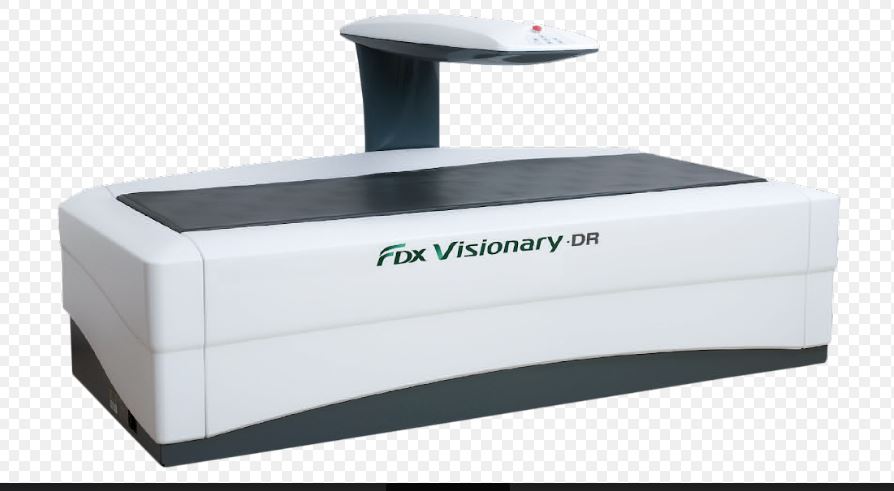
New Delhi, 19 February 2024: FUJIFILM India, a pioneer in diagnostic imaging solutions, has announced the installation of its powerful Dual-Energy X-ray Absorptiometry (DEXA) machine “FDX Visionary-DR” at the grand inauguration of the Center for Sports Injury in Delhi. The event took place on the 11th of February with the honourable dignitaries from centre alongside the presence of top sporting icons. The Center for Sports Injury is the premier sports health and wellness facility that specializes in Orthopedics, Sports Surgery, Sports Medicine, Sports Imaging, Sports Physiotherapy, Rehabilitation, Fitness and Counseling. The initiative aims to deliver high-quality and fast bone-density scans, commonly known as DEXA scans, to diagnose bone-related health conditions or evaluate the risk of developing such problems. With its innovative machinery, FUJIFILM India endeavors to make excellent diagnostic capabilities accessible to bone densitometry practitioners nationwide.
Bone-related health diseases such as osteoporosis are widespread in India, with estimates indicating that 61 million individuals suffer from this condition. Furthermore, the incidence of osteoporosis tends to occur 10 – 20 years earlier here, in comparison to Western countries. Tackling this, the bone densitometry scan serves as an effective tool to accurately predict the chances of fractures or abnormalities in the upcoming years. FUJIFILM India’s FDX Visionary-DR machine delivers a complete diagnostic experience with a wide range of applications and routine examinations for Orthopedics, Pediatrics, Lateral Spine, Morphometry and Whole Body. It features a breakthrough 3D-DXA technology that uses routine bone-mineral-density images to create 3D models of the femur, bringing in new information about bone structure for accurate diagnosis and adapted treatment procedures. Based on a 4-line, 64-element multi-array detector, the machine’s remarkable 2D Fan Beam is designed to provide the highest image resolution for a precise diagnosis and perform quick 15-second assessments per site, making it one of the most powerful solutions available in the market. Additionally, it offers advanced fat, muscle and lean medical parameters through its BODY COMPOSITION tool which is essential in sports medicine, weight management and medical practice fields.
“In India, the growing prevalence of bone-related health conditions highlights the urgent need for preventive measures in healthcare. As a leader in diagnostic imaging solutions, FUJIFILM India recognizes the critical role of early detection and timely intervention in mitigating the impact of bone-related diseases on patients and society as a whole. Thus, we are proud to introduce this FDX Visionary-DR machine in Delhi as it possesses the potential to revolutionize the bone health-related diagnostic imaging segment. Through breakthrough imaging technologies, strategic partnerships and awareness drives, our mission is to effectively diagnose, treat and manage bone health conditions and pave the way towards a healthier future for India” said, Mr. Chander Shekhar Sibal, Vice President & HOD, Healthcare Business, FUJIFILM India.
Osteoporosis occurs due to excessive bone loss, insufficient bone formation or a combination of both factors. Consequently, bones become weak, making them susceptible to fractures even from minor accidents or from everyday actions like sneezing or bumps in severe cases. Addressing this, dual-energy X-ray absorptiometry or DEXA scans measure bone loss by utilizing a minimal dose of ionizing radiation to generate images of internal body structures such as the hip, spine and forearm. It also helps with the periodic monitoring of body fitness parameters like densitometry and body composition analysis, examining the density of peripheral central body parts as well as visceral and subcutaneous fat analysis.
Dr Pushpinder – an eminent figure with over 30 years of Experience in the field of Sports Medicine, Joint Replacement Surgery & Arthroscopic Surgery (KEYHOLE SURGERY) nationally & internationally while embracing the meaningful installation said, “The alarming estimate of millions suffering from bone-related conditions like osteoporosis highlights a pressing issue which is affecting the quality of life across the nation. Also, the fact that these critical diseases tend to occur earlier in India, necessitates urgent attention to preventive care and supporting action to improve the situation. So, we take great pride in welcoming this sophisticated DEXA machine, courtesy of FUJIFILM India, at our facility to deliver reliable diagnostic services in Delhi and help communities lead a healthier life.”
FUJIFILM India reaffirms its commitment to reshaping India’s healthcare sector with the exceptional FDX Visionary-DR machine, equipping medical professionals with future-ready diagnostic technologies to provide crucial and urgent patient care.
19, Feb 2024
Pediatric Neurologist , Dr. Vykunta Raju K. N., Receives Prestigious Award for Outstanding Contribution to Child Neurology
Bengaluru, February 19, 2024: In a momentous occasion, Dr. Vykunta Raju K. N., Professor and Head of the Department at Indira Gandhi Institute of Child Health, has been honored with the Outstanding Contribution Award for Child Neurology by the Association of Child Neurology, India (AOCN). Dr. Raju, with over 15 years of dedicated service in pediatric neurology, has played a pivotal role in the diagnosis and management of rare and neurological disorders.
The accolade comes as recognition for Dr. Vykunta Raju’s tireless efforts in advancing the understanding and treatment of rare diseases, particularly in pediatric neurology. Despite the challenges faced in the field, he remains committed to improving the lives of children and families affected by these disorders. Dr. Raju has published over 200 publications focusing on original reviews and case reports on rare disorders to enhance awareness, understanding prevalence, and studying epidemiological factors in the Indian population.
While expressing his happiness for the award recognition, Dr Raju, said, “I am feeling happy to receive this award. I have been working in pediatric neurology for the past 15 years, and we have done a lot of work. Though the recognition has come a little late, I still feel very happy on getting recognized for the work I have contributed. In pediatric neurology, you will be managing a lot of rare disorders, most of the time. In earlier days, these rare diseases were neglected and treated as cerebral palsy. But now, with improvement in genetics, we are able to diagnose these disorders. Arriving at the right diagnosis was a huge challenge in the earlier days. Compared to that, diagnosis is easier, but getting proper treatment is still an issue. Treatment is not available for many disorders; and even if it is available, the cost of the medicines is very high. So, most of our patients are unable to afford these treatments.”
Speaking on Rare Disease Awareness Month (February), Dr. Raju highlighted the importance of accurate diagnosis and the need for FDA-approved treatments, addressing the difficulties faced by patients, particularly in accessing expensive medications for some disorders like Spinal Muscular Atrophy. In most of the countries, these medications are made available mostly by Government funding. So, if it’s made available in India also, it will benefit both the children and their families.
19, Feb 2024
Amblyopia – if Untreated, It Can Affect a Child’s Vision Permanently
Amblyopia is the most common vision issue that affects kids. Amblyopia (lazy eye) causes blurry vision in one eye when something affects how a child’s eyes are developing. As their brain ignores the weaker eye, that eye drifts out of position. It’s rare, but amblyopia can affect both eyes at the same time. It affects around 5% of children younger than 15 years. Eye care specialist will treat amblyopia by making child’s brain use their weaker eye to see.
This will repair and strengthen the connection between your child’s brain and both their eyes to correct the amblyopia. Most kids need amblyopic treatment at least for few months. Treatment can be done by eye patching, atropine therapy or by spectacle correction. This can include targeted education, awareness campaigns, and policy interventions.
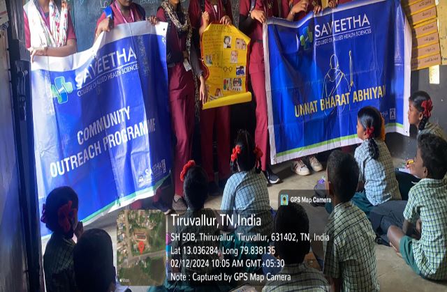
As a part of UBA and community outreach program, students and faculty of “Saveetha College of Allied Health Sciences” reached kondancherry village, Tiruvallur District, Tamil Nadu and conducted an awareness program by stating the Importance of eye screening in children to prevent amblyopia.
17, Feb 2024
Revitalizing Movement Beyond Boundaries – Venture for Innovation in Physiotherapy Practice
Telehealth and Digital Rehabilitation: Implement telehealth solutions to reach patients beyond geographical boundaries. This enables physiotherapists to provide remote consultations, monitor exercises, and offer guidance, increasing accessibility to rehabilitation services.
Virtual Reality (VR) and Augmented Reality (AR): Integrate VR and AR technologies into physiotherapy exercises to make rehabilitation more engaging and effective. This can enhance patient motivation and compliance while providing a novel approach to movement therapy.
Data-driven Personalized Treatment Plans: Utilize data analytics and wearable devices to gather real-time information about patients’ movements and progress. This data can be used to tailor personalized treatment plans, optimizing outcomes and accelerating recovery.
Robotics and Exoskeletons: Explore the use of robotics and exoskeletons in physiotherapy to assist patients in performing movements and exercises. This can be particularly beneficial for individuals with mobility challenges, providing support and enabling more intensive rehabilitation.
Gamification of Rehabilitation: Incorporate game-like elements into physiotherapy exercises to make them more enjoyable and motivating. Gamification can encourage consistent participation and adherence to rehabilitation programs, especially in the case of long-term or repetitive exercises.
Cross-disciplinary Collaboration: Foster collaboration between physiotherapists and professionals from other fields, such as technology, engineering, and design. This interdisciplinary approach can lead to the development of innovative solutions that address the complex challenges in rehabilitation.
Biomechanics and Wearable Technology: Embrace advancements in biomechanics and wearable technology to assess and monitor movement patterns. This information can guide physiotherapists in designing targeted interventions and tracking progress more accurately.
Mind-Body Integration Techniques: Integrate mindfulness and relaxation techniques into physiotherapy practices. This holistic approach considers the interconnectedness of physical and mental well-being, promoting a more comprehensive and patient-centred rehabilitation experience.
Global Knowledge Exchange: Facilitate international collaboration and knowledge exchange among physiotherapy professionals. This can involve sharing best practices, research findings, and innovative techniques to enhance the global standard of physiotherapy care.
16, Feb 2024
Living with Vertigo: Tips for a Balanced Life
Dr. Jyotirmay S Hegde, HOD & Lead Consultant ENT, Aster Whitefield Hospital, Bengaluru
You might have experienced vertigo when you look down from a tall height and suddenly feel dizzy, with a sensation like butterflies in your stomach. This is called vertigo and it is a fairly common issue that most people face. This happens because your brain gets mixed signals about your body’s position, especially when you’re on the edge of a tall building or looking down from a height. This conflicting information can make you feel unsteady or dizzy, which is a common symptom of vertigo.
Vertigo covers a range of feelings such as feeling faint, light-headed, or unsteady, along with the sensation of spinning or motion while you’re still. It’s often connected to problems in the inner ear or brain. You don’t have to be at a high place to experience vertigo; it can happen even on a flat surface. You may feel like you’re spinning, tilting, swaying, or moving, which can affect your daily life and make it hard to keep your balance.
Vertigo can have various causes, and although there isn’t clear evidence directly linking it to genetics, some conditions that contribute to vertigo may have genetic components. Inner ear problems like Meniere’s disease or vestibular neuritis, a tendency to have migraines, aging, head injuries, certain medications, vestibular disorders, dehydration, and anemia can all play a role in causing vertigo. While genetic factors might make someone more prone to conditions related to vertigo, it’s usually a mix of genetic, environmental, and lifestyle factors that lead to its occurrence.
It’s important for everyone, especially women, to take care of their inner ear health to prevent vertigo. This means avoiding loud noises, protecting ears from infections, managing stress, and having a balanced diet with enough vitamin D and magnesium. Women may be particularly affected due to hormonal changes during pregnancy or menopause, which can influence inner ear function and make them more prone to vertigo. Additionally, conditions like osteoporosis, which are more common in women, can indirectly impact inner ear health. However, vertigo is a concern for both genders and is part of overall health maintenance and prevention strategies for everyone.
To manage vertigo at home, there are several self-care tips you can follow. These include making sure you drink enough water to stay hydrated, avoiding sudden movements that can trigger vertigo episodes, getting enough rest, and practicing relaxation techniques such as deep breathing or meditation. Specific physical therapy exercises like the Epley maneuver can also be helpful in relieving symptoms. It’s important to adopt a healthy lifestyle overall, which includes managing stress, getting adequate sleep, staying hydrated, and eating a balanced diet. These lifestyle changes can contribute to your overall well-being and may help reduce the impact of vertigo.
Lifestyle choices like stress, not getting enough sleep, being dehydrated, consuming too much caffeine or alcohol, and having poor eating habits can make vertigo worse or trigger it. Certain dietary factors such as dehydration, and consuming too much salt or caffeine, can make symptoms worse by affecting fluid balance, blood pressure, and how the inner ear works. While these lifestyle factors may not directly cause vertigo, they can make someone more likely to have episodes or make existing symptoms worse. Not getting enough sleep can affect how well your brain works, including its ability to maintain balance. If you don’t drink enough fluids, it can lower your blood volume and change your blood pressure, which affects the flow of oxygen to your brain and inner ear, leading to balance issues. Caffeine and alcohol can both affect blood flow and disrupt how the inner ear works, which can contribute to vertigo symptoms. Not having enough essential nutrients in your diet, like vitamin D and magnesium, can affect your overall health, including the health of your inner ear. A lack of these nutrients may contribute to conditions that cause vertigo.
Engaging in vestibular rehabilitation exercises can be beneficial for reducing vertigo symptoms by aiding in the adaptation and compensation of the vestibular system. It’s important for individuals experiencing vertigo to steer clear of activities that involve rapid head movements or positions that can trigger episodes. The outlook for vertigo varies depending on its root cause, with some cases being manageable or treatable while others may persist chronically or recur.
In summary, vertigo is a complex condition influenced by problems in the inner ear and lifestyle factors. Conditions like Meniere’s disease and migraines can contribute to its occurrence, and although genetic connections are being investigated, lifestyle aspects such as stress and diet also have a significant impact. Taking a comprehensive approach, including personalized guidance from ENT specialists or doctors, is essential for managing vertigo and enhancing overall well-being.
—
16, Feb 2024
Six safest position to hold a new born baby
By Dr.N.Senthil Kumar, MPT PhD
Mr. Srikanth. V MPT (Student)
Saveetha College of Physiotherapy, SIMATS, Thandalam, Chennai.
1. Cradle hold:
Baby parallel to your chest level, slide your hand from baby bottom up to support the head and neck. Kindly place your baby’s head into the crook of your elbow. Now move your hand from the neck to the bottom and hips. Bring your baby close to you.
2. Shoulder hold:
- This is one of the most natural holds for your new born. Here is how you should do it:
With the baby’s body parallel to yours, lift her to your shoulder height. Rest her head on your shoulder so that she can look behind you. Support her head and neck with one hand, and the bottom with another hand. This position allows your baby to hear your heartbeat.
3. Belly hold:
- Your baby will surely enjoy this baby hold. Here is how to handle the newborn:
Lay your baby with stomach facing down on one of your forearms with head up over the elbow. Let her feet land on either side of your hand, at an angle closer to the ground level. Lay your other hand across the baby’s back to make her feel secure. This baby positioning technique can be helpful for burping. Gently stroke the baby’s back to help release any trapped gas.
4. Hip hold:
- You can hold your baby’s hips once she gains complete control over her head and neck. So, try this position when your baby crosses three months. Here is how you can do this: Face your baby outward and make her sit on your hip bones. You should wrap your arm around her waist. Your baby will be able to look at things around her comfortably.
5. Face to face hold:
- This hold will let you interact with your baby. Here is how you can try the hold perfectly: Support your baby’s head and neck with one hand. Offer support to her bottom with another hand. Now hold the baby just below the chest facing you. Enjoy making her smile and have fun interacting with her.
6. Football hold:
- This hold also serves as a feeding position; you can use it while standing or sitting. Here is how you should do it: Support your baby’s neck and head with your hand, and the rest of her back with your same forearm. Adjust the baby’s head and neck with the other hand. Make the baby curl towards your body side, with her legs extended behind. Draw your little one closer to your chest. Use the other free hand for offering extra support to the head or feed the baby.
16, Feb 2024
BSE Listed Nutricircle Launches Health Supplement, My45Nutrients
NOIDA, February 16, 2024: Nutricircle, a BSE-listed R&D and science-based company, introduces another health supplement, My45Nutrients, under its nutraceutical category. My45Nutrients- Daily Dose of Goodness, made of plant extracts, tablets are designed to provide a comprehensive range of essential nutrients, including 8 vitamins, 29 amino acids + bio-nutrients, and 8 minerals. These nutrients are formulated to support various aspects of overall well-being.
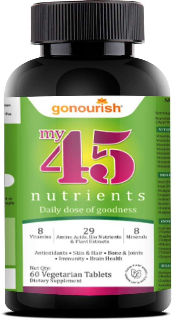
Enriched with essential vitamins, minerals, antioxidants, amino acids, bio-nutrients, and natural plant extracts, My45 Nutrients supplement is designed to bolster immune function and promote overall vitality. This comprehensive formula protects cells from damage, contributes to the normal function of the immune system, helps support daily energy needs, increases the ability
to handle stress, controls anxiety, expert support for hair, contributes to the normal functioning of the nervous system & heart, and protects the skin. It is anti-inflammatory, repairs, and maintains, cartilage, bones, and teeth, and revitalizes the system to be energetic.
“The newest introduction to our gamut of health supplements, My45Nutrients stands out with a unique formula that includes essential nutrients sourced from plants, which helps in boosting immunity and bolsters the overall well-being of the body. With lifestyle issues seeing an upward graph, this product aims to address daily health needs, ensuring that everyone receives a well– rounded selection of vital nutrients to maintain and promote overall health said Hitesh M Patel, Promoter & Director, Nutricircle.
My45Nutrients is densely packed with all the necessary nutrients the body needs to support better health and wellness. This product comprises plant-based ingredients such as Korean Ginseng root extract, grape seed extract, PhyllanthusEmblica dried fruit extract (Amla), Moringa, stevia, Curcuma longa root powder (Turmeric), Ginger, Garlic, Tulsi and BacopaMonnieri. Nutricircle has tied up with Aghub, ICRISAT, IIMR, Nutrihub, and NIFTEM for product development and R&D.
My45Nutrients will be available on various online platforms as well as offline stores in cities- Mumbai, Gujarat, and Hyderabad, in the initial phase.
16, Feb 2024
Are you at risk of Preeclampsia
Are you an expecting Mom? While the joy of impending motherhood is certainly exciting, there’s important information to be aware of as you embark on this incredible journey. Preeclampsia is a syndrome that manifests during pregnancy, characterized by elevated blood pressure and signs of organ damage, typically affecting the heart, liver, and kidneys. This condition is usually identified after the 20th week of pregnancy and can impact both the expectant mother and the developing foetus.
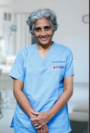
Preeclampsia can be tricky to detect and one should be extra cautious of the symptoms and should not ignore even a small sign of it. Talking openly with the doctor will certainly help to manage the condition positively to ensure maximum protection for the baby and mother, says Dr. Prathima Reddy, Director & Lead Consultant, Department of Obstetrics and Gynaecology, SPARSH Hospital, Bangalore.
#Signs and Symptoms:
o Watch out for increased blood pressure (hypertension) often with headaches and visual disturbance.
o There can be swelling of hands and face or sudden weight gain.
o Continuous nausea or vomiting may imply severe preeclampsia.
#Risk Factors:
o These include having a family history of preeclampsia.
o Take heed if you are pregnant with twins or more, as well as if you are suffering from a previously existing illness (high blood pressure, diabetes, kidney disease and the like).
#Complications:
o The two incidences that come along with it are premature birth and low birth weight of the baby
o In severe instances, it may progress to eclampsia, marked by seizures, thereby posing serious risks to the mother’s life.
#Regular Prenatal Check-ups:
o Regular check-ups will help the physician monitor your blood pressure and preeclampsia symptoms during your visits.
o If preeclampsia is detected early chances are higher for both mother and baby to have a better prognosis.
#Lifestyle Modifications:
o To be in good health, it is necessary to have a balanced diet, especially fruits and vegetables and whole-grain products.
o Engage in physical activity under your physician’s supervision. This could involve following a recommended exercise routine tailored by your doctor.
o Stressed out? Try breathing deeply or signing up for prenatal yoga classes.
#Know When to Seek Help:
o Do not wait, call your doctor immediately if you notice any symptoms that could be worrying
o Preeclampsia can only be effectively managed through emergency medical attention.
#The Power of Support:
Have open conversations with your healthcare team, partner, and family regarding your concerns and the experiences you’ve encountered in this matter.
When a woman is aware of preeclampsia, she is empowered to take proactive steps to ensure the safe and healthy birth of her baby. In the exciting journey of motherhood, the path ahead is one of upward progress. Knowledge is key, so stay informed, remain vigilant, implement lifestyle adjustments, and approach pregnancy with confidence.
15, Feb 2024
Supporting the Diagnosis of Bone-related Diseases, FUJIFILM India Installs an Advanced DEXA Machine in Delhi
New Delhi, 15 February 2024: FUJIFILM India, a pioneer in diagnostic imaging solutions, has announced the installation of its powerful Dual-Energy X-ray Absorptiometry (DEXA) machine “FDX Visionary-DR” at the grand inauguration of the Center for Sports Injury in Delhi. The event took place on the 11th of February with the honourable dignitaries from centre alongside the presence of top sporting icons. The Center for Sports Injury is the premier sports health and wellness facility that specializes in Orthopedics, Sports Surgery, Sports Medicine, Sports Imaging, Sports Physiotherapy, Rehabilitation, Fitness and Counseling. The initiative aims to deliver high-quality and fast bone-density scans, commonly known as DEXA scans, to diagnose bone-related health conditions or evaluate the risk of developing such problems. With its innovative machinery, FUJIFILM India endeavors to make excellent diagnostic capabilities accessible to bone densitometry practitioners nationwide.
Bone-related health diseases such as osteoporosis are widespread in India, with estimates indicating that 61 million individuals suffer from this condition. Furthermore, the incidence of osteoporosis tends to occur 10 – 20 years earlier here, in comparison to Western countries. Tackling this, the bone densitometry scan serves as an effective tool to accurately predict the chances of fractures or abnormalities in the upcoming years. FUJIFILM India’s FDX Visionary-DR machine delivers a complete diagnostic experience with a wide range of applications and routine examinations for Orthopedics, Pediatrics, Lateral Spine, Morphometry and Whole Body. It features a breakthrough 3D-DXA technology that uses routine bone-mineral-density images to create 3D models of the femur, bringing in new information about bone structure for accurate diagnosis and adapted treatment procedures. Based on a 4-line, 64-element multi-array detector, the machine’s remarkable 2D Fan Beam is designed to provide the highest image resolution for a precise diagnosis and perform quick 15-second assessments per site, making it one of the most powerful solutions available in the market. Additionally, it offers advanced fat, muscle and lean medical parameters through its BODY COMPOSITION tool which is essential in sports medicine, weight management and medical practice fields.
“In India, the growing prevalence of bone-related health conditions highlights the urgent need for preventive measures in healthcare. As a leader in diagnostic imaging solutions, FUJIFILM India recognizes the critical role of early detection and timely intervention in mitigating the impact of bone-related diseases on patients and society as a whole. Thus, we are proud to introduce this FDX Visionary-DR machine in Delhi as it possesses the potential to revolutionize the bone health-related diagnostic imaging segment. Through breakthrough imaging technologies, strategic partnerships and awareness drives, our mission is to effectively diagnose, treat and manage bone health conditions and pave the way towards a healthier future for India” said, Mr. Chander Shekhar Sibal, Vice President & HOD, Healthcare Business, FUJIFILM India.
Osteoporosis occurs due to excessive bone loss, insufficient bone formation or a combination of both factors. Consequently, bones become weak, making them susceptible to fractures even from minor accidents or from everyday actions like sneezing or bumps in severe cases. Addressing this, dual-energy X-ray absorptiometry or DEXA scans measure bone loss by utilizing a minimal dose of ionizing radiation to generate images of internal body structures such as the hip, spine and forearm. It also helps with the periodic monitoring of body fitness parameters like densitometry and body composition analysis, examining the density of peripheral central body parts as well as visceral and subcutaneous fat analysis.
Dr Pushpinder – an eminent figure with over 30 years of Experience in the field of Sports Medicine, Joint Replacement Surgery & Arthroscopic Surgery (KEYHOLE SURGERY) nationally & internationally while embracing the meaningful installation said, “The alarming estimate of millions suffering from bone-related conditions like osteoporosis highlights a pressing issue which is affecting the quality of life across the nation. Also, the fact that these critical diseases tend to occur earlier in India, necessitates urgent attention to preventive care and supporting action to improve the situation. So, we take great pride in welcoming this sophisticated DEXA machine, courtesy of FUJIFILM India, at our facility to deliver reliable diagnostic services in Delhi and help communities lead a healthier life.”
FUJIFILM India reaffirms its commitment to reshaping India’s healthcare sector with the exceptional FDX Visionary-DR machine, equipping medical professionals with future-ready diagnostic technologies to provide crucial and urgent patient care.